6HK5 image
Deposition Date 2018-09-05
Release Date 2019-03-27
Last Version Date 2024-11-13
Entry Detail
PDB ID:
6HK5
Title:
X-ray structure of a truncated mutant of the metallochaperone CooJ with a high-affinity nickel-binding site
Biological Source:
Source Organism:
Method Details:
Experimental Method:
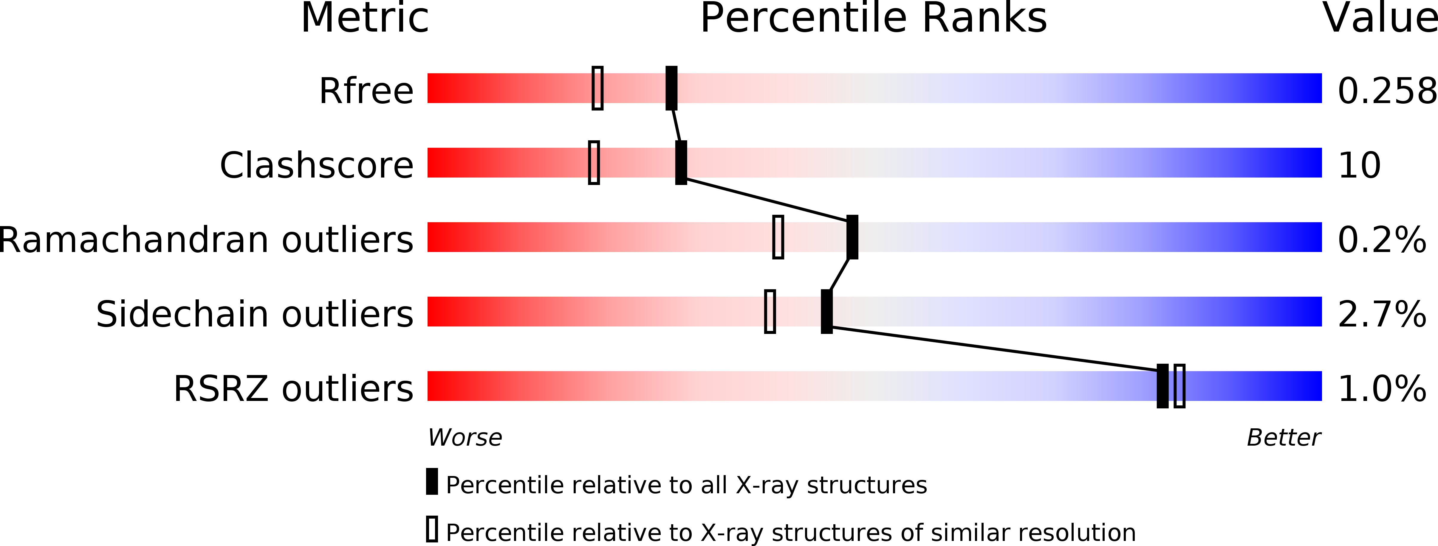
Resolution:
2.04 Å
R-Value Free:
0.25
R-Value Work:
0.19
R-Value Observed:
0.20
Space Group:
P 1 21 1
Macromolecular Entities
Polymer Type:polypeptide(L)
Molecule:CooJ
Gene (Uniprot):cooJ
Chain IDs:A (auth: B), B (auth: C), C (auth: E), D (auth: G), E (auth: A), F (auth: D), G (auth: F), H
Chain Length:68
Number of Molecules:8
Biological Source:Rhodospirillum rubrum
Modified Residue
Compound ID Chain ID Parent Comp ID Details 2D Image
MSE A MET modified residue
Primary Citation
The carbon monoxide dehydrogenase accessory protein CooJ is a histidine-rich multidomain dimer containing an unexpected Ni(II)-binding site.
J.Biol.Chem. 294 7601 7614 (2019)
PMID: 30858174 DOI: 10.1074/jbc.RA119.008011

Abstact

Activation of nickel enzymes requires specific accessory proteins organized in multiprotein complexes controlling metal transfer to the active site. Histidine-rich clusters are generally present in at least one of the metallochaperones involved in nickel delivery. The maturation of carbon monoxide dehydrogenase in the proteobacterium Rhodospirillum rubrum requires three accessory proteins, CooC, CooT, and CooJ, dedicated to nickel insertion into the active site, a distorted [NiFe3S4] cluster coordinated to an iron site. Previously, CooJ from R. rubrum (RrCooJ) has been described as a nickel chaperone with 16 histidines and 2 cysteines at its C terminus. Here, the X-ray structure of a truncated version of RrCooJ, combined with small-angle X-ray scattering data and a modeling study of the full-length protein, revealed a homodimer comprising a coiled coil with two independent and highly flexible His tails. Using isothermal calorimetry, we characterized several metal-binding sites (four per dimer) involving the His-rich motifs and having similar metal affinity (KD = 1.6 μm). Remarkably, biophysical approaches, site-directed mutagenesis, and X-ray crystallography uncovered an additional nickel-binding site at the dimer interface, which binds Ni(II) with an affinity of 380 nm Although RrCooJ was initially thought to be a unique protein, a proteome database search identified at least 46 bacterial CooJ homologs. These homologs all possess two spatially separated nickel-binding motifs: a variable C-terminal histidine tail and a strictly conserved H(W/F)X2HX3H motif, identified in this study, suggesting a dual function for CooJ both as a nickel chaperone and as a nickel storage protein.

Legend

Protein

Chemical

Disease

Primary Citation of related structures