
Deposition Date
2018-09-04
Release Date
2019-10-09
Last Version Date
2024-01-17
Entry Detail
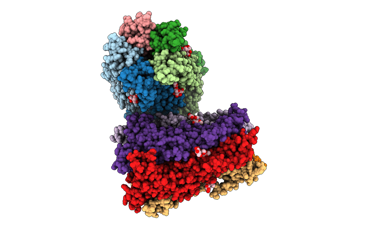
PDB ID:
6HK0
Keywords:
Title:
X-ray structure of a pentameric ligand gated ion channel from Erwinia chrysanthemi (ELIC) F16'S pore mutant (F247S) with alternate M4 conformation.
Biological Source:
Source Organism:
Dickeya chrysanthemi (Taxon ID: 556)
Host Organism:
Method Details:
Experimental Method:
Resolution:
3.45 Å
R-Value Free:
0.26
R-Value Work:
0.22
R-Value Observed:
0.22
Space Group:
P 1 21 1
