Deposition Date
2018-09-04
Release Date
2019-10-09
Last Version Date
2024-10-23
Entry Detail
PDB ID:
6HJX
Keywords:
Title:
X-ray structure of a pentameric ligand gated ion channel from Erwinia chrysanthemi (ELIC) 7'C pore mutant (L238C) in complex with nanobody 72
Biological Source:
Source Organism(s):
Dickeya chrysanthemi (Taxon ID: 556)
Lama glama (Taxon ID: 9844)
Lama glama (Taxon ID: 9844)
Expression System(s):
Method Details:
Experimental Method:
Resolution:
2.50 Å
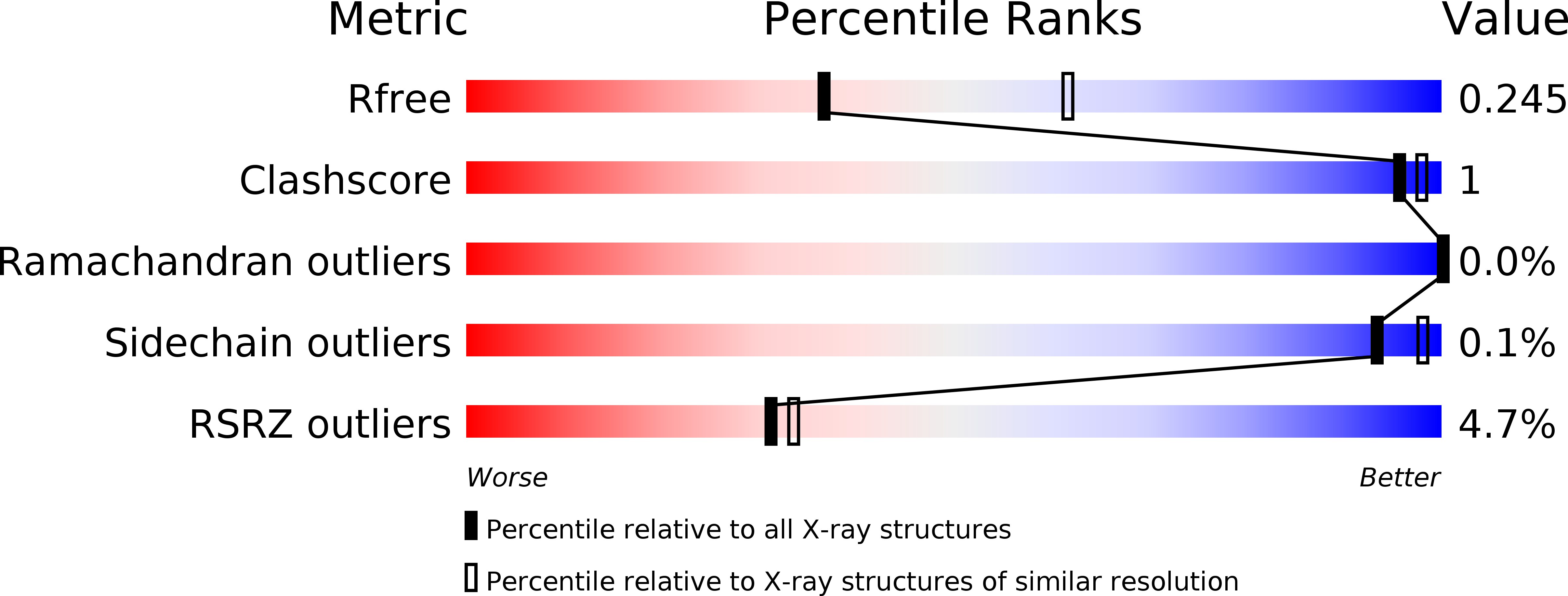
R-Value Free:
0.24
R-Value Work:
0.20
R-Value Observed:
0.20
Space Group:
P 1 21 1