6HIT image
Deposition Date 2018-08-30
Release Date 2019-08-21
Last Version Date 2024-11-13
Entry Detail
PDB ID:
6HIT
Title:
The crystal structure of haemoglobin from Atlantic cod
Biological Source:
Source Organism:
Gadus morhua (Taxon ID: 8049)
Method Details:
Experimental Method:
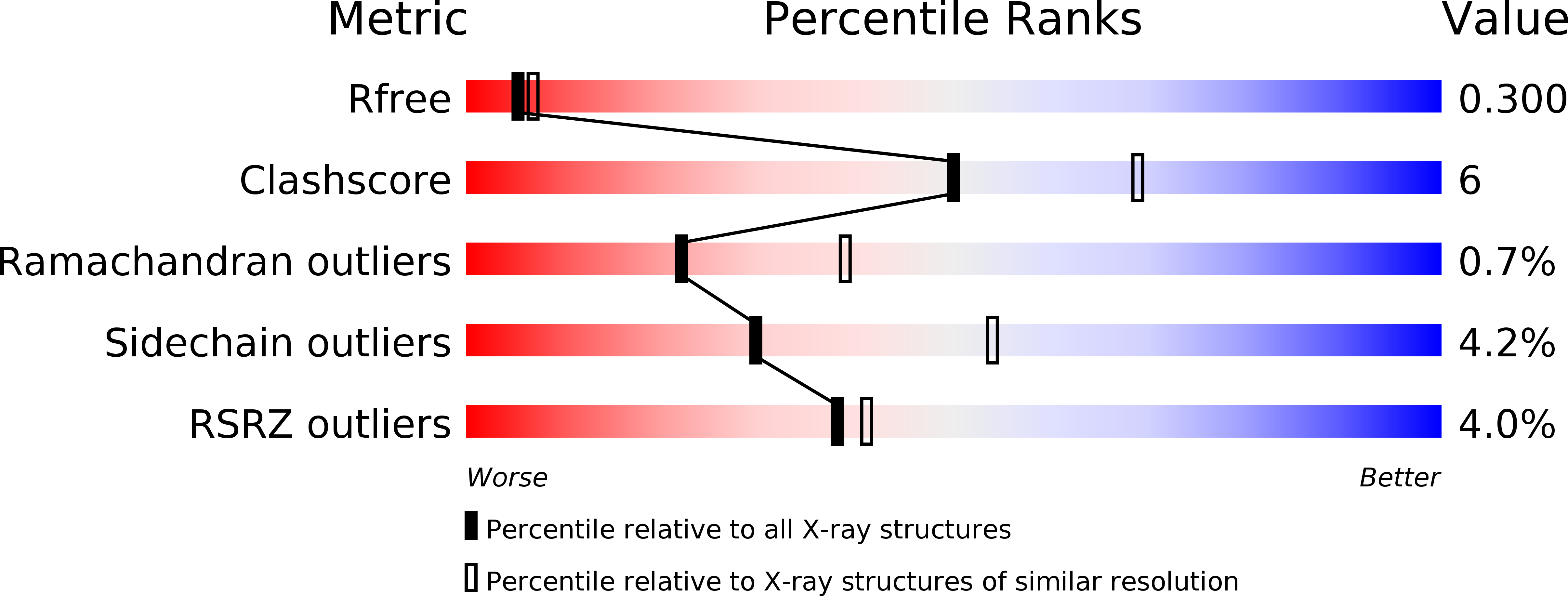
Resolution:
2.50 Å
R-Value Free:
0.30
R-Value Work:
0.23
R-Value Observed:
0.23
Space Group:
P 21 21 21
Macromolecular Entities
Polymer Type:polypeptide(L)
Molecule:Hemoglobin alpha 2 chain
Chain IDs:A, C, E, G
Chain Length:143
Number of Molecules:4
Biological Source:Gadus morhua
Polymer Type:polypeptide(L)
Molecule:Hemoglobin beta 4 chain
Gene (Uniprot):HbB4
Chain IDs:B, D, F, H
Chain Length:145
Number of Molecules:4
Biological Source:Gadus morhua
Ligand Molecules
Primary Citation
The crystal structure of haemoglobin from Atlantic cod.
Acta Crystallogr.,Sect.F 75 537 542 (2019)
PMID: 31397324 DOI: 10.1107/S2053230X1900904X

Abstact

The crystal structure of haemoglobin from Atlantic cod has been solved to 2.54 Å resolution. The structure consists of two tetramers in the crystallographic asymmetric unit. The structure of haemoglobin obtained from one individual cod suggests polymorphism in the tetrameric assembly.

Legend

Protein

Chemical

Disease

Primary Citation of related structures