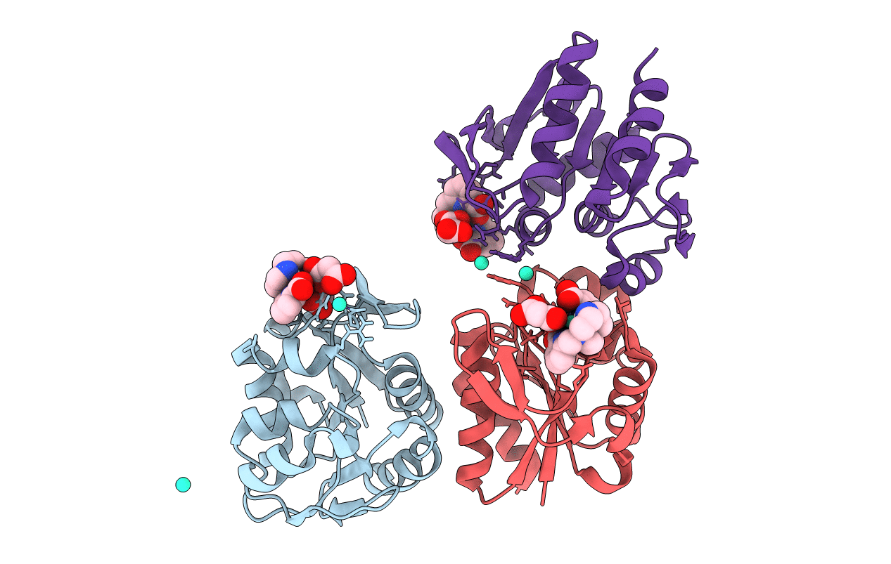
6HF6 image
Deposition Date 2018-08-21
Release Date 2019-06-19
Last Version Date 2024-05-15
Entry Detail
PDB ID:
6HF6
Keywords:
Title:
Crystal structure of the Protease 1 (E29A,E60A,E80A) from Pyrococcus horikoshii co-crystallized with Tb-Xo4.
Biological Source:
Method Details:
Experimental Method:
Resolution:
2.00 Å
R-Value Free:
0.18
R-Value Work:
0.16
R-Value Observed:
0.16
Space Group:
P 41 21 2
Macromolecular Entities
Structures with similar UniProt ID
Protein Blast
Polymer Type:polypeptide(L)
Molecule:Deglycase PH1704
Gene (Uniprot):PH1704
Chain IDs:A, B, C
Chain Length:166
Number of Molecules:3
Biological Source:Pyrococcus horikoshii (strain ATCC 700860 / DSM 12428 / JCM 9974 / NBRC 100139 / OT-3)
Primary Citation
Protein crystal structure determination with the crystallophore, a nucleating and phasing agent.
J.Appl.Crystallogr. 52 722 731 (2019)
PMID: 31396026 DOI: 10.1107/S1600576719006381

Abstact

Obtaining crystals and solving the phase problem remain major hurdles encountered by bio-crystallographers in their race to obtain new high-quality structures. Both issues can be overcome by the crystallophore, Tb-Xo4, a lanthanide-based molecular complex with unique nucleating and phasing properties. This article presents examples of new crystallization conditions induced by the presence of Tb-Xo4. These new crystalline forms bypass crystal defects often encountered by crystallographers, such as low-resolution diffracting samples or crystals with twinning. Thanks to Tb-Xo4's high phasing power, the structure determination process is greatly facilitated and can be extended to serial crystallography approaches.

Legend

Protein

Chemical

Disease

Primary Citation of related structures