Deposition Date
2018-08-16
Release Date
2018-12-05
Last Version Date
2024-10-23
Entry Detail
PDB ID:
6HCS
Keywords:
Title:
Crystal structure of CaM-peptide complex containing AzF at position 108
Biological Source:
Source Organism(s):
Homo sapiens (Taxon ID: 9606)
Rattus norvegicus (Taxon ID: 10116)
Rattus norvegicus (Taxon ID: 10116)
Expression System(s):
Method Details:
Experimental Method:
Resolution:
2.00 Å
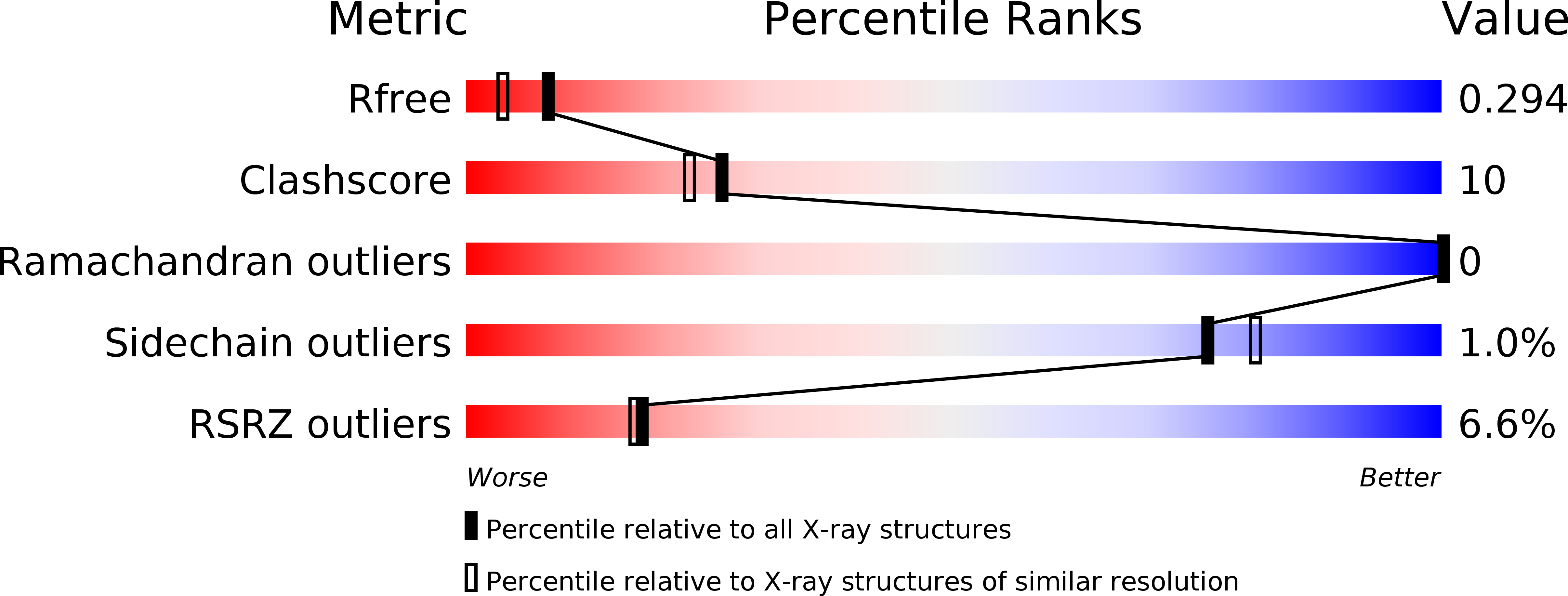
R-Value Free:
0.29
R-Value Work:
0.26
R-Value Observed:
0.26
Space Group:
P 1 21 1