6HCI image
Deposition Date 2018-08-15
Release Date 2019-08-28
Last Version Date 2024-05-01
Entry Detail
PDB ID:
6HCI
Keywords:
Title:
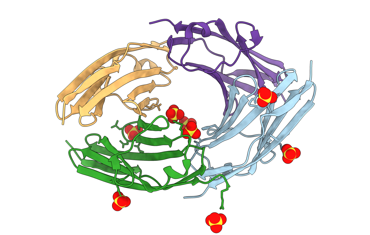
Crystal structure of titin M3 domain
Biological Source:
Source Organism:
Homo sapiens (Taxon ID: 9606)
Host Organism:
Method Details:
Experimental Method:
Resolution:
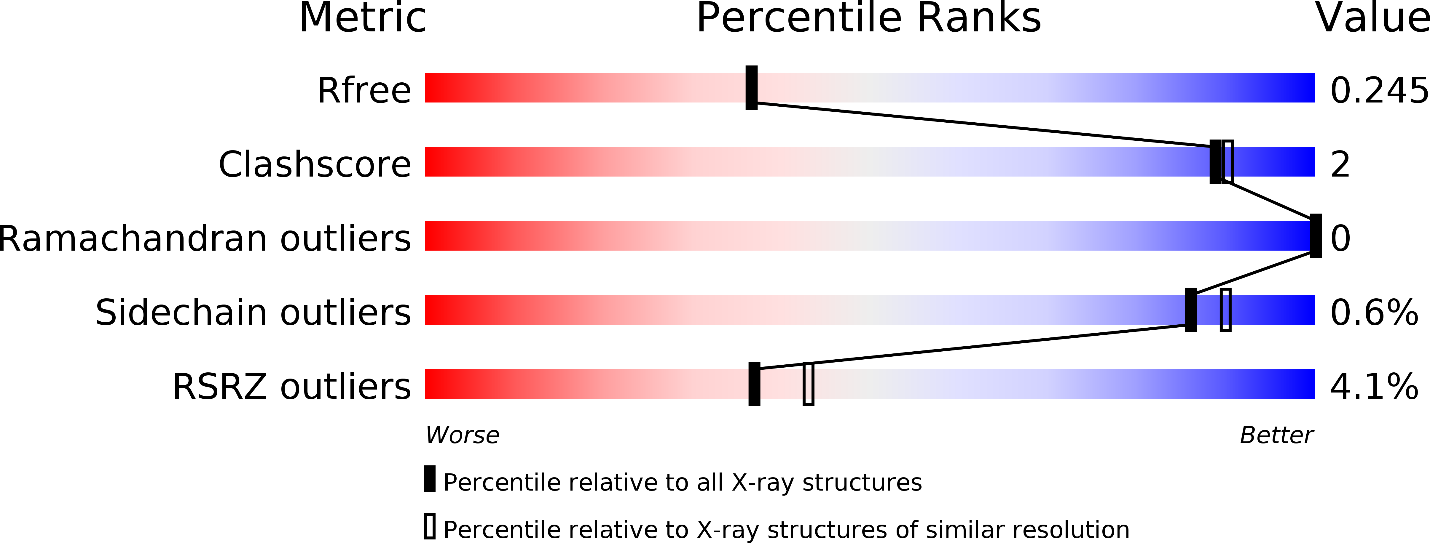
2.12 Å
R-Value Free:
0.24
R-Value Work:
0.19
R-Value Observed:
0.19
Space Group:
P 32 2 1
Macromolecular Entities
Structures with similar UniProt ID
Protein Blast
Polymer Type:polypeptide(L)
Molecule:Titin
Gene (Uniprot):TTN
Chain IDs:A, B, C, D
Chain Length:105
Number of Molecules:4
Biological Source:Homo sapiens
Ligand Molecules
Primary Citation
Structural diversity in the atomic resolution 3D fingerprint of the titin M-band segment.
Plos One 14 e0226693 e0226693 (2019)
PMID: 31856237 DOI: 10.1371/journal.pone.0226693

Abstact

In striated muscles, molecular filaments are largely composed of long protein chains with extensive arrays of identically folded domains, referred to as "beads-on-a-string". It remains a largely unresolved question how these domains have developed a unique molecular profile such that each carries out a distinct function without false-positive readout. This study focuses on the M-band segment of the sarcomeric protein titin, which comprises ten identically folded immunoglobulin domains. Comparative analysis of high-resolution structures of six of these domains ‒ M1, M3, M4, M5, M7, and M10 ‒ reveals considerable structural diversity within three distinct loops and a non-conserved pattern of exposed cysteines. Our data allow to structurally interpreting distinct pathological readouts that result from titinopathy-associated variants. Our findings support general principles that could be used to identify individual structural/functional profiles of hundreds of identically folded protein domains within the sarcomere and other densely crowded cellular environments.

Legend

Protein

Chemical

Disease

Primary Citation of related structures