Deposition Date
1998-09-10
Release Date
1998-09-16
Last Version Date
2023-09-20
Entry Detail
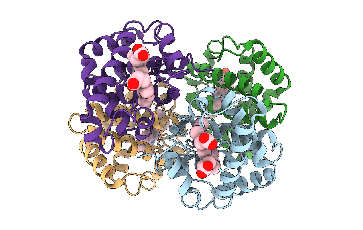
PDB ID:
6HBW
Keywords:
Title:
Crystal structure of deoxy-human hemoglobin beta6 glu->trp
Biological Source:
Source Organism(s):
Homo sapiens (Taxon ID: 9606)
Expression System(s):
Method Details:
Experimental Method:
Resolution:
2.00 Å
R-Value Free:
0.26
R-Value Work:
0.19
R-Value Observed:
0.19
Space Group:
P 21 21 21