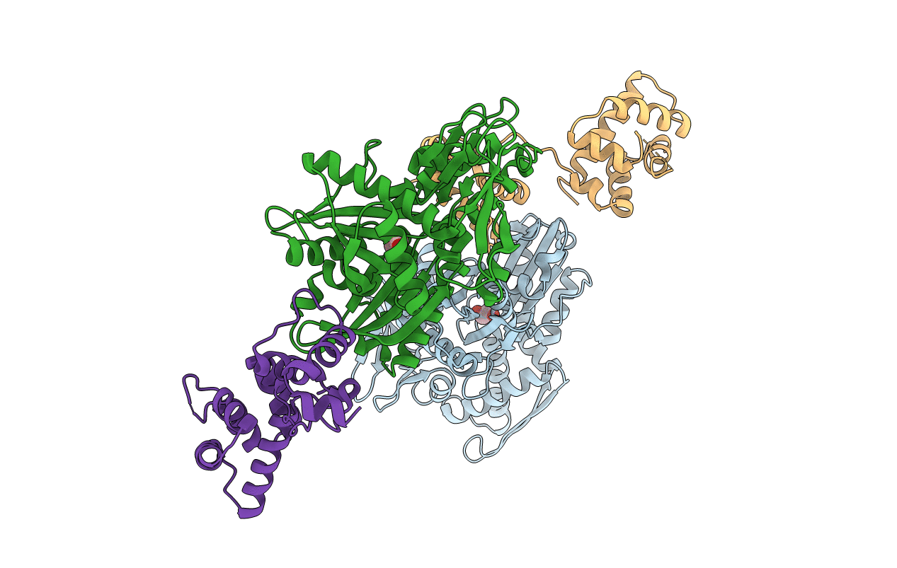
6HA7 image
Entry Detail
PDB ID:
6HA7
Keywords:
Title:
Crystal structure of the BiP NBD and MANF complex
Biological Source:
Host Organism:
PDB Version:
Deposition Date:
2018-08-07
Release Date:
2019-02-06
Method Details:
Experimental Method:
Resolution:
2.49 Å
R-Value Free:
0.25
R-Value Work:
0.22
R-Value Observed:
0.22
Space Group:
P 1
Macromolecular Entities
Polymer Type:polypeptide(L)
Description:Endoplasmic reticulum chaperone BiP
Chain IDs:A, C (auth: B)
Chain Length:387
Number of Molecules:2
Biological Source:Cricetulus griseus
Polymer Type:polypeptide(L)
Description:Mesencephalic astrocyte-derived neurotrophic factor
Chain IDs:B (auth: C), D
Chain Length:158
Number of Molecules:2
Biological Source:Mus musculus
Ligand Molecules
Primary Citation
MANF antagonizes nucleotide exchange by the endoplasmic reticulum chaperone BiP.
Nat Commun 10 541 541 (2019)
PMID: 30710085 DOI: 10.1038/s41467-019-08450-4

Abstact

Despite its known role as a secreted neuroprotectant, much of the mesencephalic astrocyte-derived neurotrophic factor (MANF) is retained in the endoplasmic reticulum (ER) of producer cells. There, by unknown mechanisms, MANF plays a role in protein folding homeostasis in complex with the ER-localized Hsp70 chaperone BiP. Here we report that the SAF-A/B, Acinus, and PIAS (SAP) domain of MANF selectively associates with the nucleotide binding domain (NBD) of ADP-bound BiP. In crystal structures the SAP domain engages the cleft between NBD subdomains Ia and IIa, stabilizing the ADP-bound conformation and clashing with the interdomain linker that occupies this site in ATP-bound BiP. MANF inhibits both ADP release from BiP and ATP binding to BiP, and thereby client release. Cells lacking MANF have fewer ER stress-induced BiP-containing high molecular weight complexes. These findings suggest that MANF contributes to protein folding homeostasis as a nucleotide exchange inhibitor that stabilizes certain BiP-client complexes.

Legend

Protein

Chemical

Disease

Primary Citation of related structures