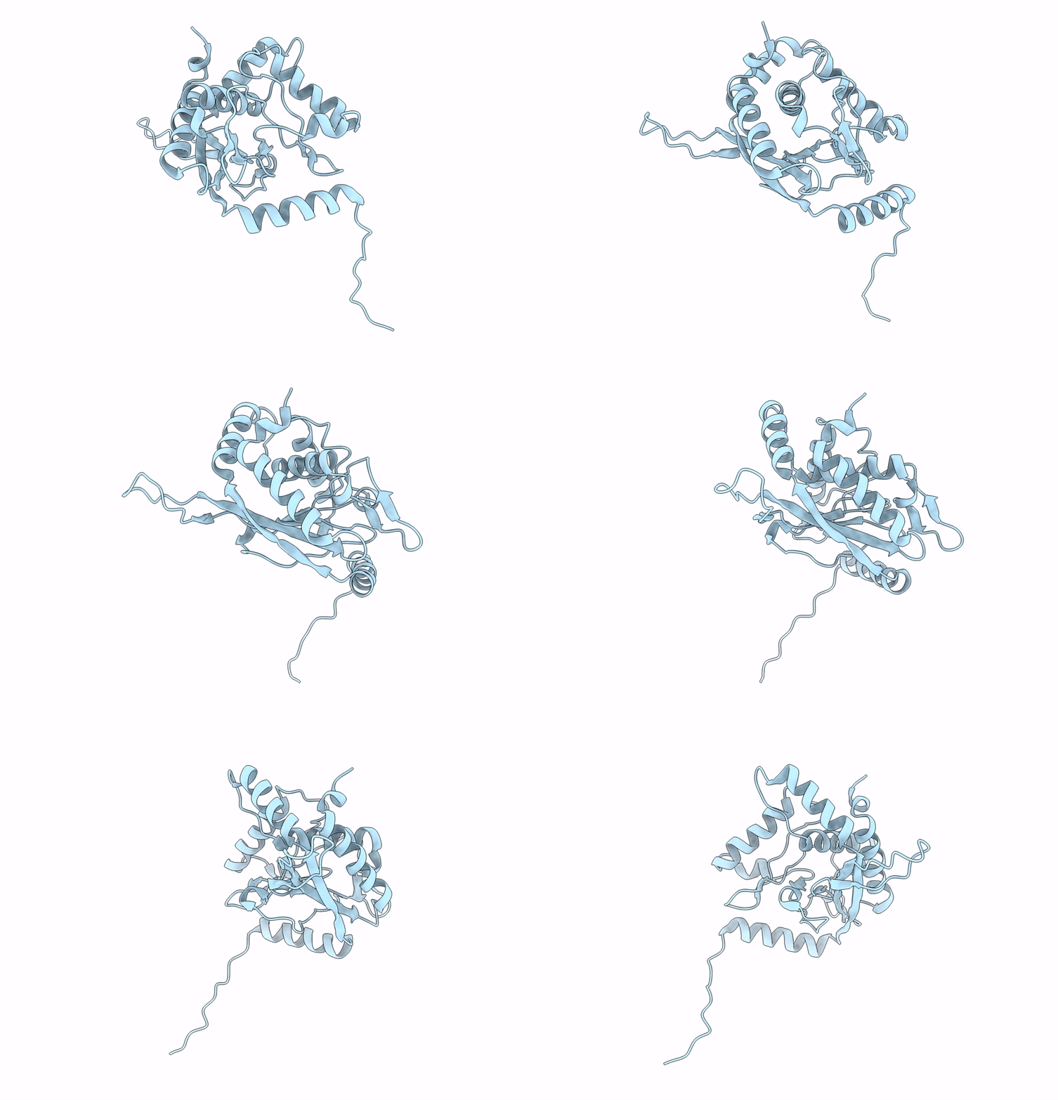
6H55 image
Deposition Date 2018-07-23
Release Date 2019-06-05
Last Version Date 2024-05-15
Entry Detail
PDB ID:
6H55
Keywords:
Title:
core of the human pyruvate dehydrogenase (E2)
Biological Source:
Source Organism:
Homo sapiens (Taxon ID: 9606)
Host Organism:
Method Details:
Experimental Method:
Resolution:
6.00 Å
Aggregation State:
PARTICLE
Reconstruction Method:
SINGLE PARTICLE
Macromolecular Entities
Polymer Type:polypeptide(L)
Molecule:Dihydrolipoyllysine-residue acetyltransferase component of pyruvate dehydrogenase complex, mitochondrial
Gene (Uniprot):DLAT
Chain IDs:A
Chain Length:647
Number of Molecules:1
Biological Source:Homo sapiens
Ligand Molecules
Primary Citation
Structural and Functional Analyses of the Human PDH Complex Suggest a "Division-of-Labor" Mechanism by Local E1 and E3 Clusters.
Structure 27 1124 1136.e4 (2019)
PMID: 31130485 DOI: 10.1016/j.str.2019.04.009

Abstact

The pseudo-atomic structural model of human pyruvate dehydrogenase complex (PDHc) core composed of full-length E2 and E3BP components, calculated from our cryoelectron microscopy-derived density maps at 6-Å resolution, is similar to those of prokaryotic E2 structures. The spatial organization of human PDHc components as evidenced by negative-staining electron microscopy and native mass spectrometry is not homogeneous, and entails the unanticipated formation of local clusters of E1:E2 and E3BP:E3 complexes. Such uneven, clustered organization translates into specific duties for E1-E2 clusters (oxidative decarboxylation and acetyl transfer) and E3BP-E3 clusters (regeneration of reduced lipoamide) corresponding to half-reactions of the PDHc catalytic cycle. The addition of substrate coenzyme A modulates the conformational landscape of PDHc, in particular of the lipoyl domains, extending the postulated multiple random coupling mechanism. The conformational and associated chemical landscapes of PDHc are thus not determined entirely stochastically, but are restrained and channeled through an asymmetric architecture and further modulated by substrate binding.

Legend

Protein

Chemical

Disease

Primary Citation of related structures