Deposition Date
2018-07-06
Release Date
2018-12-12
Last Version Date
2024-01-17
Entry Detail
PDB ID:
6H07
Keywords:
Title:
X-ray structure of Lactobacillus brevis alcohol dehydrogenase
Biological Source:
Source Organism(s):
Lactobacillus brevis (Taxon ID: 1580)
Expression System(s):
Method Details:
Experimental Method:
Resolution:
1.48 Å
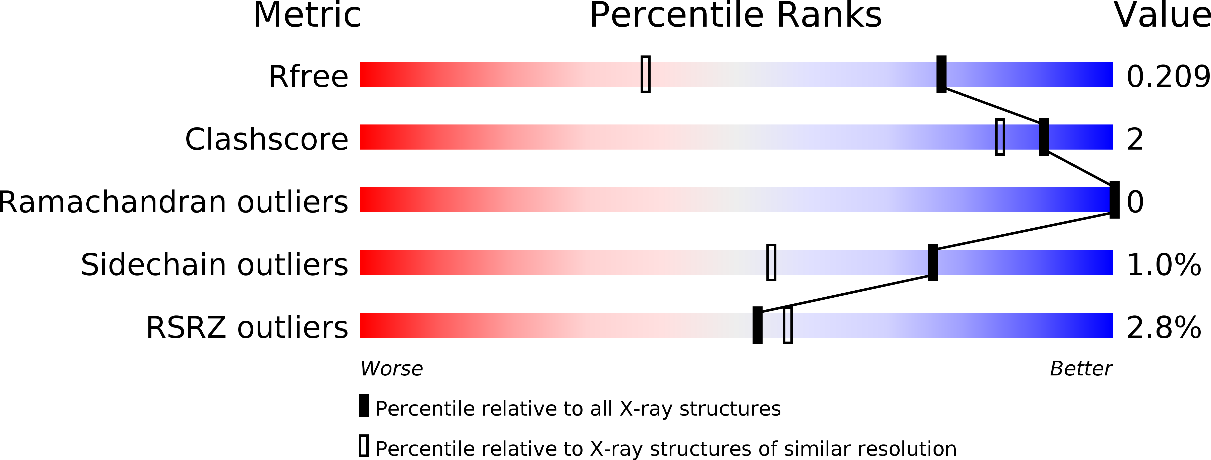
R-Value Free:
0.20
R-Value Work:
0.18
R-Value Observed:
0.18
Space Group:
P 21 2 21