6H03 image
Deposition Date 2018-07-06
Release Date 2018-12-19
Last Version Date 2024-11-13
Entry Detail
PDB ID:
6H03
Keywords:
Title:
OPEN CONFORMATION OF THE MEMBRANE ATTACK COMPLEX
Biological Source:
Source Organism:
Homo sapiens (Taxon ID: 9606)
Method Details:
Experimental Method:
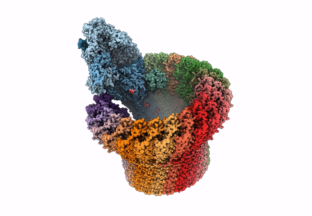
Resolution:
5.60 Å
Aggregation State:
PARTICLE
Reconstruction Method:
SINGLE PARTICLE
Macromolecular Entities
Polymer Type:polypeptide(L)
Molecule:Complement C5,Complement C5
Gene (Uniprot):C5
Chain IDs:A
Chain Length:1580
Number of Molecules:1
Biological Source:Homo sapiens
Polymer Type:polypeptide(L)
Molecule:Complement component C6
Gene (Uniprot):C6
Chain IDs:F (auth: B)
Chain Length:913
Number of Molecules:1
Biological Source:Homo sapiens
Polymer Type:polypeptide(L)
Molecule:Complement component C8 beta chain
Gene (Uniprot):C8B
Chain IDs:B (auth: C)
Chain Length:537
Number of Molecules:1
Biological Source:Homo sapiens
Polymer Type:polypeptide(L)
Molecule:Complement component C7
Gene (Uniprot):C7
Chain IDs:C (auth: D)
Chain Length:821
Number of Molecules:1
Biological Source:Homo sapiens
Polymer Type:polypeptide(L)
Molecule:Complement component C8 gamma chain
Gene (Uniprot):C8G
Chain IDs:D (auth: E)
Chain Length:182
Number of Molecules:1
Biological Source:Homo sapiens
Polymer Type:polypeptide(L)
Molecule:Complement component C8 alpha chain
Gene (Uniprot):C8A
Chain IDs:E (auth: F)
Chain Length:554
Number of Molecules:1
Biological Source:Homo sapiens
Polymer Type:polypeptide(L)
Molecule:Complement component C9
Gene (Uniprot):C9
Chain IDs:G, H (auth: P), I (auth: H), J (auth: I), K (auth: J), L (auth: K), M (auth: L), N (auth: M), O (auth: N), P (auth: O), Q, R, S, T, U, V, W, X
Chain Length:538
Number of Molecules:18
Biological Source:Homo sapiens
Ligand Molecules
Primary Citation
CryoEM reveals how the complement membrane attack complex ruptures lipid bilayers.
Nat Commun 9 5316 5316 (2018)
PMID: 30552328 DOI: 10.1038/s41467-018-07653-5

Abstact

The membrane attack complex (MAC) is one of the immune system's first responders. Complement proteins assemble on target membranes to form pores that lyse pathogens and impact tissue homeostasis of self-cells. How MAC disrupts the membrane barrier remains unclear. Here we use electron cryo-microscopy and flicker spectroscopy to show that MAC interacts with lipid bilayers in two distinct ways. Whereas C6 and C7 associate with the outer leaflet and reduce the energy for membrane bending, C8 and C9 traverse the bilayer increasing membrane rigidity. CryoEM reconstructions reveal plasticity of the MAC pore and demonstrate how C5b6 acts as a platform, directing assembly of a giant β-barrel whose structure is supported by a glycan scaffold. Our work provides a structural basis for understanding how β-pore forming proteins breach the membrane and reveals a mechanism for how MAC kills pathogens and regulates cell functions.

Legend

Protein

Chemical

Disease

Primary Citation of related structures