Deposition Date
2018-06-28
Release Date
2018-07-25
Last Version Date
2024-05-15
Entry Detail
Biological Source:
Source Organism(s):
Expression System(s):
Method Details:
Experimental Method:
Resolution:
2.50 Å
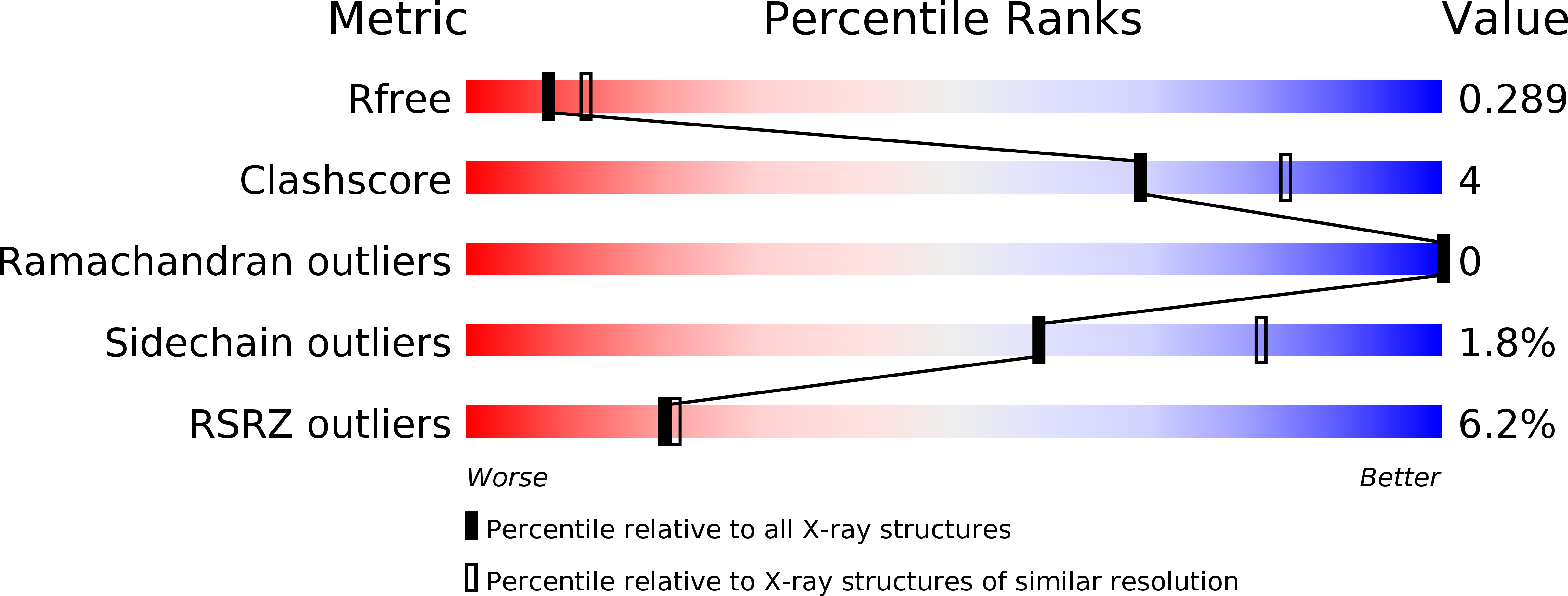
R-Value Free:
0.28
R-Value Work:
0.23
R-Value Observed:
0.24
Space Group:
P 21 21 21