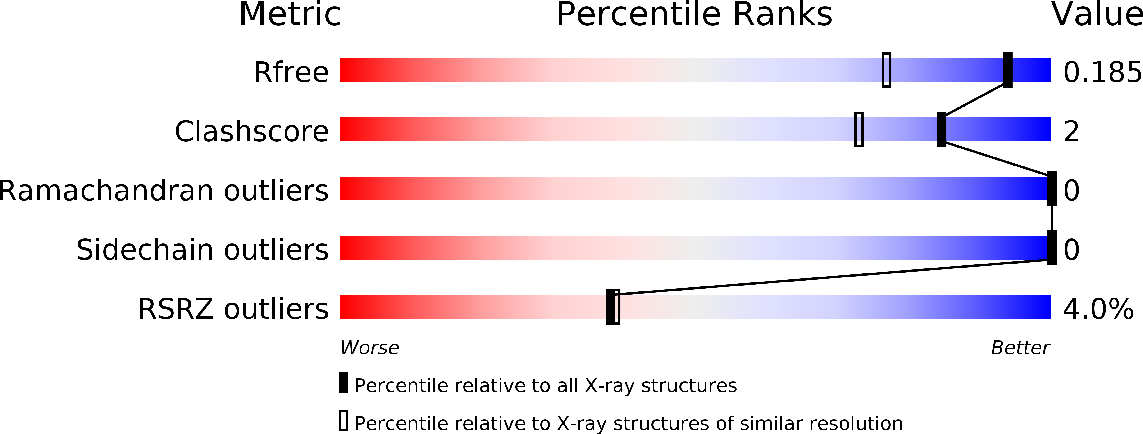
6GXJ image
Deposition Date 2018-06-27
Release Date 2019-02-06
Last Version Date 2024-01-17
Entry Detail
PDB ID:
6GXJ
Keywords:
Title:
X-ray structure of DiRu-1-encapsulated Apoferritin
Biological Source:
Source Organism:
Equus caballus (Taxon ID: 9796)
Method Details:
Experimental Method:
Resolution:
1.43 Å
R-Value Free:
0.19
R-Value Work:
0.17
R-Value Observed:
0.17
Space Group:
F 4 3 2
Macromolecular Entities
Polymer Type:polypeptide(L)
Molecule:Ferritin light chain
Gene (Uniprot):FTL
Chain IDs:A
Chain Length:174
Number of Molecules:1
Biological Source:Equus caballus
Primary Citation
Encapsulation of the Dinuclear Trithiolato-Bridged Arene Ruthenium Complex Diruthenium-1 in an Apoferritin Nanocage: Structure and Cytotoxicity.
ChemMedChem 14 594 602 (2019)
PMID: 30674089 DOI: 10.1002/cmdc.201800805

Abstact

The effects of encapsulating the cytotoxic dinuclear trithiolato-bridged arene ruthenium complex [(η6 -p-MeC6 H4 iPr)2 Ru2 (μ2 -S-p-C6 H4 tBu)3 ]Cl (DiRu-1) within the apoferritin (AFt) nanocage were investigated. The DiRu-1-AFt nanocarrier was characterized by UV/Vis spectroscopy, ICP-MS, CD and X-ray crystallography. In contrast to previously reported Au- and Pt-based drug-loaded AFt carriers, we found no evidence of direct interactions between DiRu-1 and AFt. DiRu-1-AFt is cytotoxic toward immortalized murine BALB/c-3T3 fibroblasts transformed with SV40 virus (SVT2) and human epidermoid carcinoma A431 malignant cells, and exhibits moderate selectivity for these cancer cells over normal BALB/c-3T3 cells. DiRu-1-AFt triggers the production of reactive oxygen species, depolarization of mitochondrial membrane potential, and induces cell death via p53-mediated apoptosis. Comparison between our data and previous results suggests that the presence of specific interactions between a metal-based drug and AFt within the protein cage is not essential for drug encapsulation.

Legend

Protein

Chemical

Disease

Primary Citation of related structures