
Deposition Date
2018-06-07
Release Date
2018-12-19
Last Version Date
2024-01-17
Entry Detail
PDB ID:
6GQH
Keywords:
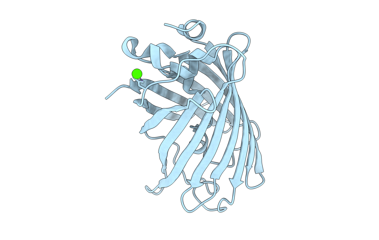
Title:
Structure of GFPmut2 crystallized at pH 8.5 and transferred to pH 6
Biological Source:
Source Organism(s):
Aequorea victoria (Taxon ID: 6100)
Expression System(s):
Method Details:
Experimental Method:
Resolution:
2.40 Å
R-Value Free:
0.23
R-Value Work:
0.19
R-Value Observed:
0.19
Space Group:
P 21 21 21
