6GQC image
Entry Detail
PDB ID:
6GQC
Keywords:
Title:
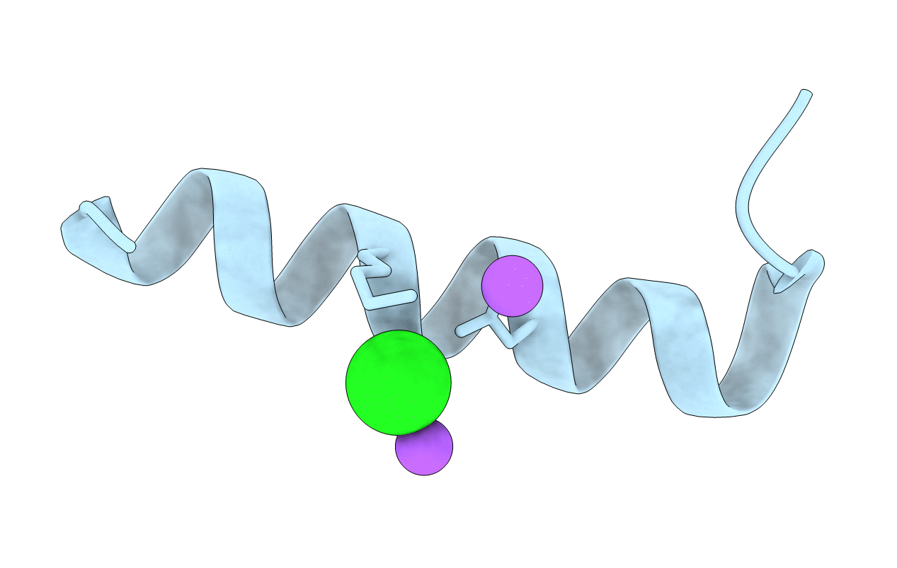
Crystal Structure of the PSMalpha3 Peptide Mutant G16A Forming Cross-Alpha Amyloid-like Fibril
Biological Source:
PDB Version:
Deposition Date:
2018-06-07
Release Date:
2019-06-19
Method Details:
Experimental Method:
Resolution:
1.40 Å
R-Value Free:
0.19
R-Value Work:
0.15
R-Value Observed:
0.15
Space Group:
C 1 2 1
Macromolecular Entities
Polymer Type:polypeptide(L)
Description:Phenol-soluble modulin alpha 3 peptide
Mutations:G16A
Chain IDs:A
Chain Length:22
Number of Molecules:1
Biological Source: Staphylococcus aureus subsp. aureus NCTC 8325
Primary Citation
Staphylococcus aureus PSM alpha 3 Cross-alpha Fibril Polymorphism and Determinants of Cytotoxicity.
Structure 28 301 313.e6 (2020)
PMID: 31918959 DOI: 10.1016/j.str.2019.12.006

Abstact

The phenol-soluble modulin (PSM) peptide family, secreted by Staphylococcus aureus, performs various virulence activities, some mediated by the formation of amyloid fibrils of diverse architectures. Specifically, PSMα1 and PSMα4 structure the S. aureus biofilm by assembling into robust cross-β amyloid fibrils. PSMα3, the most cytotoxic member of the family, assembles into cross-α fibrils in which α helices stack into tightly mated sheets, mimicking the cross-β architecture. Here we demonstrate that massive T cell deformation and death are linked with PSMα3 aggregation and co-localization with cell membranes. Our extensive mutagenesis analyses support the role of positive charges, and especially Lys17, in interactions with the membrane and suggest their regulation by inter- and intra-helical electrostatic interactions within the cross-α fibril. We hypothesize that PSMα3 cytotoxicity is governed by the ability to form cross-α fibrils and involves a dynamic process of co-aggregation with the cell membrane, rupturing it.

Legend

Protein

Chemical

Disease

Primary Citation of related structures