6GNZ image
Entry Detail
PDB ID:
6GNZ
Title:
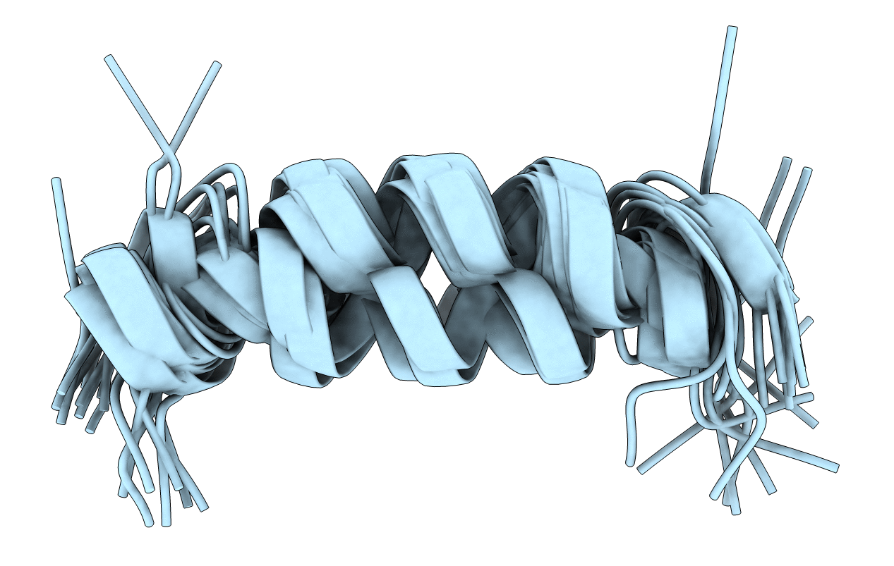
Plantaricin S-a in 100 mM DPC micelles. This is the alpha part of the bacteriocin plantaricin S.
Biological Source:
Source Organism:
PDB Version:
Deposition Date:
2018-06-01
Release Date:
2019-03-06
Method Details:
Experimental Method:
Conformers Calculated:
100
Conformers Submitted:
20
Selection Criteria:
structures with the lowest energy
Macromolecular Entities
Polymer Type:polypeptide(L)
Description:Plantaricin S alpha protein
Chain IDs:A
Chain Length:27
Number of Molecules:1
Biological Source:Lactobacillus plantarum
Ligand Molecules
Primary Citation
NMR structures and mutational analysis of the two peptides constituting the bacteriocin plantaricin S.
Sci Rep 9 2333 2333 (2019)
PMID: 30787405 DOI: 10.1038/s41598-019-38518-6

Abstact

The structure of the individual peptides of the two-peptide bacteriocin plantaricin S, an antimicrobial peptide produced by a Lactobacillus plantarum strain, has been determined in DPC micelles. The two peptides of plantaricin S, Pls-α and Pls-β, form an α-helix from and including residue 8 to 24 with a less structured region around residue 16-19 and an amphiphilic α-helix from and including residue 7 to 23, respectively. Activity assays on single amino acid-substituted GxxxG and GxxxG-like motifs show that substituting the Ser and Gly residues in the G9xxxG13 motif in Pls-α and the S17xxxG21 motif in Pls-β reduced or drastically reduced the antimicrobial activity. The two-peptide bacteriocin muricidin contains GxxxG-like motifs at similar positions and displays 40-50% amino acid identity with plantaricin S. Activity assays of combinations of the peptides that constitute the bacteriocins plantaricin S and muricidin show that some combinations are highly active. Furthermore, sequence alignments show that the motifs important for plantaricin S activity align with identical motifs in muricidin. Based on sequence comparison and activity assays, a membrane-inserted model of plantaricin S in which the two peptides are oriented antiparallel relative to each other and where the GxxxG and GxxxG-like motifs important for activity come close in space, is proposed.

Legend

Protein

Chemical

Disease

Primary Citation of related structures