6GMN image
Deposition Date 2018-05-27
Release Date 2018-08-08
Last Version Date 2024-11-20
Entry Detail
PDB ID:
6GMN
Keywords:
Title:
pVHL:EloB:EloC in complex with methyl 4H-furo[3,2-b]pyrrole-5-carboxylate
Biological Source:
Source Organism:
Homo sapiens (Taxon ID: 9606)
Host Organism:
Method Details:
Experimental Method:
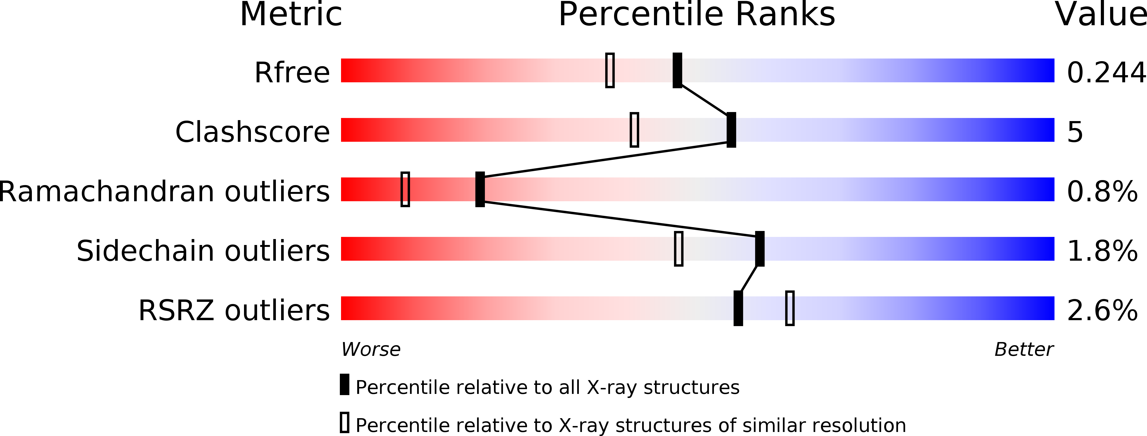
Resolution:
1.94 Å
R-Value Free:
0.24
R-Value Work:
0.20
R-Value Observed:
0.20
Space Group:
P 41 2 2
Macromolecular Entities
Structures with similar UniProt ID
Protein Blast
Polymer Type:polypeptide(L)
Molecule:Elongin-B
Gene (Uniprot):ELOB
Chain IDs:A
Chain Length:104
Number of Molecules:1
Biological Source:Homo sapiens
Structures with similar UniProt ID
Protein Blast
Polymer Type:polypeptide(L)
Molecule:Elongin-C
Gene (Uniprot):ELOC
Chain IDs:B, E, H, K
Chain Length:97
Number of Molecules:4
Biological Source:Homo sapiens
Structures with similar UniProt ID
Protein Blast
Polymer Type:polypeptide(L)
Molecule:von Hippel-Lindau disease tumor suppressor
Gene (Uniprot):VHL
Chain IDs:C, F, L
Chain Length:162
Number of Molecules:3
Biological Source:Homo sapiens
Structures with similar UniProt ID
Protein Blast
Polymer Type:polypeptide(L)
Molecule:Elongin-B
Gene (Uniprot):ELOB
Chain IDs:D, G, J
Chain Length:104
Number of Molecules:3
Biological Source:Homo sapiens
Structures with similar UniProt ID
Protein Blast
Polymer Type:polypeptide(L)
Molecule:von Hippel-Lindau disease tumor suppressor
Gene (Uniprot):VHL
Chain IDs:I
Chain Length:162
Number of Molecules:1
Biological Source:Homo sapiens
Modified Residue
Compound ID Chain ID Parent Comp ID Details 2D Image
CAS A CYS modified residue
Primary Citation
Surface Probing by Fragment-Based Screening and Computational Methods Identifies Ligandable Pockets on the von Hippel-Lindau (VHL) E3 Ubiquitin Ligase.
J. Med. Chem. 61 7387 7393 (2018)
PMID: 30040896 DOI: 10.1021/acs.jmedchem.8b00842

Abstact

Beyond the targeting of E3 ubiquitin ligases to inhibit protein homeostasis, E3 ligase binders can be repurposed as targeted protein degraders (PROTACs or molecular glues). We sought to identify new binders of the VHL E3 ligase by biophysical fragment-based screening followed by X-ray crystallographic soaking. We identified fragments binding at the ElonginC:Cullin2 interface and a new cryptic pocket in VHL, along with other potential ligandable sites predicted computationally and found to bind solvent molecules in crystal structures. The elucidated interactions provide starting points for future ligand development.

Legend

Protein

Chemical

Disease

Primary Citation of related structures
Feedback Form
Name
Email
Institute
Feedback