Deposition Date
2018-05-23
Release Date
2018-06-13
Last Version Date
2024-10-16
Entry Detail
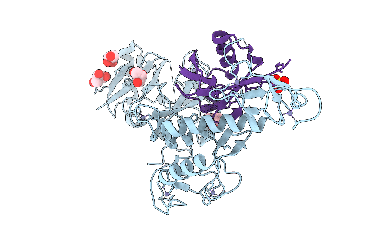
Biological Source:
Source Organism(s):
Homo sapiens (Taxon ID: 9606)
Expression System(s):
Method Details:
Experimental Method:
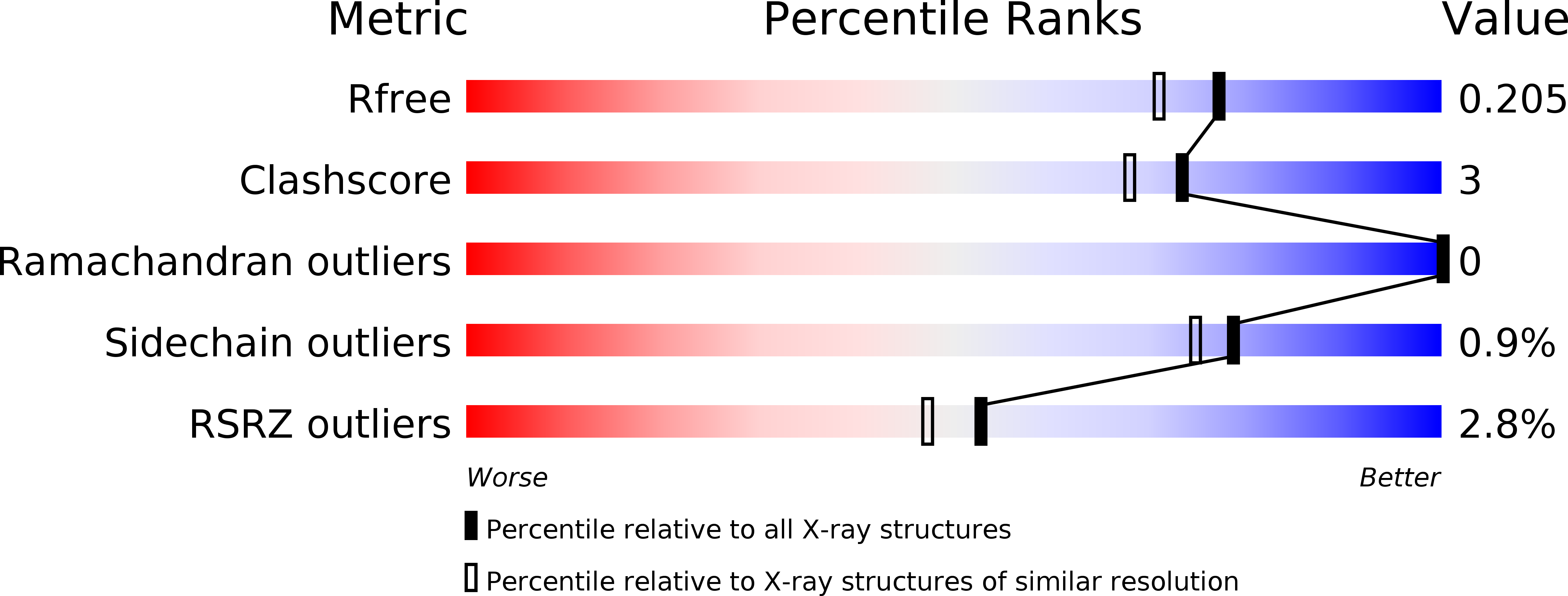
Resolution:
1.80 Å
R-Value Free:
0.20
R-Value Work:
0.17
R-Value Observed:
0.18
Space Group:
P 32 2 1