6GL2 image
Entry Detail
PDB ID:
6GL2
Keywords:
Title:
Structure of ZgEngAGH5_4 wild type at 1.2 Angstrom resolution
Biological Source:
Source Organism:
PDB Version:
Deposition Date:
2018-05-22
Release Date:
2018-10-31
Method Details:
Experimental Method:
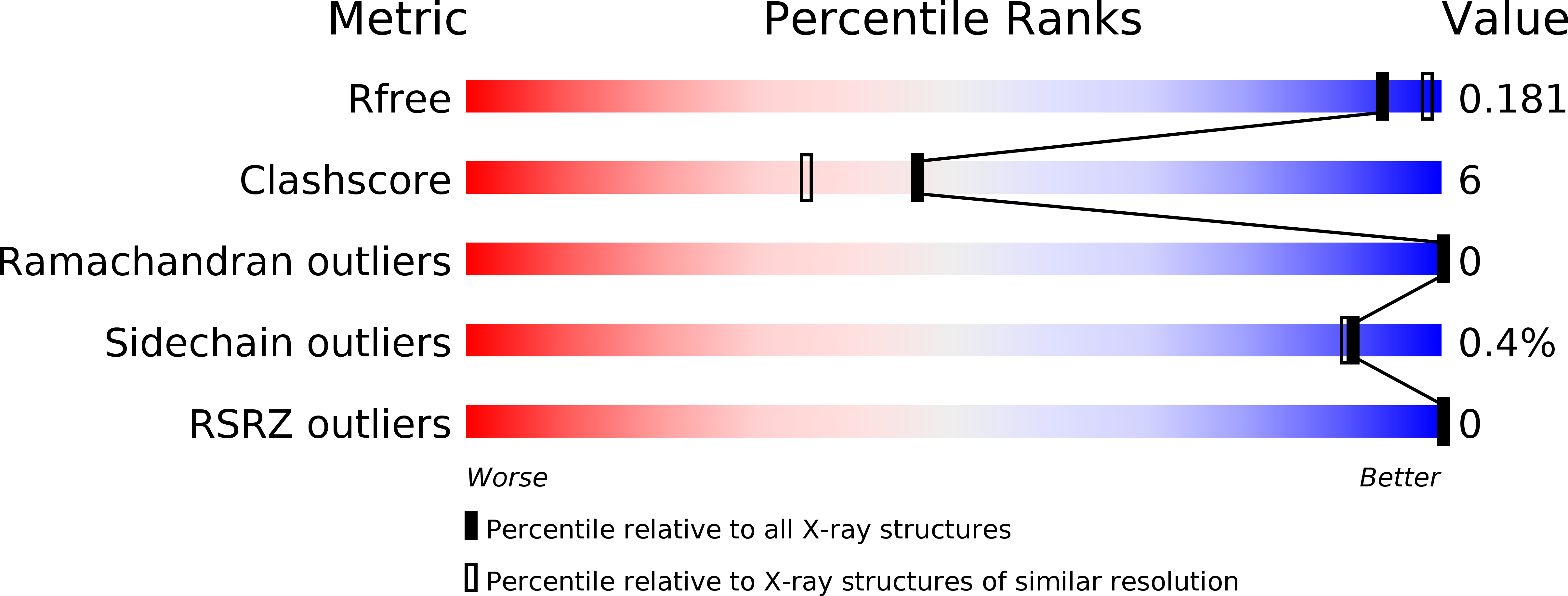
Resolution:
1.96 Å
R-Value Free:
0.20
R-Value Work:
0.17
R-Value Observed:
0.17
Space Group:
P 1 21 1
Macromolecular Entities
Polymer Type:polypeptide(L)
Description:Endoglucanase, family GH5
Chain IDs:A
Chain Length:337
Number of Molecules:1
Biological Source:Zobellia galactanivorans
Ligand Molecules
Primary Citation
The laterally acquired GH5ZgEngAGH5_4from the marine bacteriumZobellia galactanivoransis dedicated to hemicellulose hydrolysis.
Biochem. J. 475 3609 3628 (2018)
PMID: 30341165 DOI: 10.1042/BCJ20180486

Abstact

Cell walls of marine macroalgae are composed of diverse polysaccharides that provide abundant carbon sources for marine heterotrophic bacteria. Among them, Zobellia galactanivorans is considered as a model for studying algae-bacteria interactions. The degradation of typical algal polysaccharides, such as agars or alginate, has been intensively studied in this model bacterium, but the catabolism of plant-like polysaccharides is essentially uncharacterized. Here, we identify a polysaccharide utilization locus in the genome of Z. galactanivorans, induced by laminarin (β-1,3-glucans), and containing a putative GH5 subfamily 4 (GH5_4) enzyme, currently annotated as a endoglucanase (ZgEngAGH5_4). A phylogenetic analysis indicates that ZgEngAGH5_4 was laterally acquired from an ancestral Actinobacteria We performed the biochemical and structural characterization of ZgEngAGH5_4 and demonstrated that this GH5 is, in fact, an endo-β-glucanase, most active on mixed-linked glucan (MLG). Although ZgEngAGH5_4 and GH16 lichenases both hydrolyze MLG, these two types of enzymes release different series of oligosaccharides. Structural analyses of ZgEngAGH5_4 reveal that all the amino acid residues involved in the catalytic triad and in the negative glucose-binding subsites are conserved, when compared with the closest relative, the cellulase EngD from Clostridium cellulovorans, and some other GH5s. In contrast, the positive glucose-binding subsites of ZgEngAGH5_4 are different and this could explain the preference for MLG, with respect to cellulose or laminarin. Molecular dynamics computer simulations using different hexaoses reveal that the specificity for MLG occurs through the +1 and +2 subsites of the binding pocket that display the most important differences when compared with the structures of other GH5_4 enzymes.

Legend

Protein

Chemical

Disease

Primary Citation of related structures