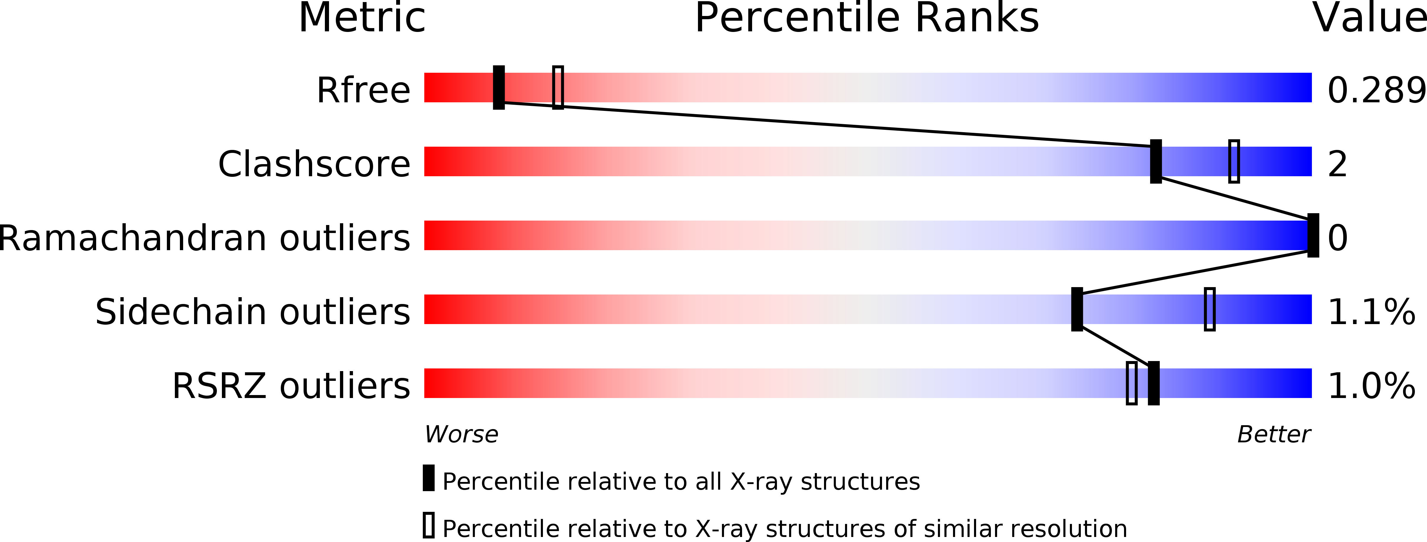
6GKF image
Deposition Date 2018-05-20
Release Date 2018-10-17
Last Version Date 2024-11-06
Entry Detail
PDB ID:
6GKF
Title:
Structure of 14-3-3 gamma in complex with caspase-2 14-3-3 binding motif Ser139
Biological Source:
Source Organism:
Homo sapiens (Taxon ID: 9606)
Method Details:
Experimental Method:
Resolution:
2.60 Å
R-Value Free:
0.28
R-Value Work:
0.24
R-Value Observed:
0.24
Space Group:
P 41 21 2
Macromolecular Entities
Structures with similar UniProt ID
Protein Blast
Polymer Type:polypeptide(L)
Molecule:14-3-3 protein gamma
Gene (Uniprot):YWHAG
Mutagens:S235Stop
Chain IDs:A, B (auth: C), C (auth: E), D (auth: G), E (auth: B), F (auth: D), G (auth: F), H
Chain Length:234
Number of Molecules:8
Biological Source:Homo sapiens
Structures with similar UniProt ID
Protein Blast
Polymer Type:polypeptide(L)
Molecule:Caspase-2
Chain IDs:I, J, K, L, M, N, O, P
Chain Length:8
Number of Molecules:8
Biological Source:Homo sapiens
Primary Citation
14-3-3 protein masks the nuclear localization sequence of caspase-2.
FEBS J. 285 4196 4213 (2018)
PMID: 30281929 DOI: 10.1111/febs.14670

Abstact

Caspase-2 is an apical protease responsible for the proteolysis of cellular substrates directly involved in mediating apoptotic signaling cascades. Caspase-2 activation is inhibited by phosphorylation followed by binding to the scaffolding protein 14-3-3, which recognizes two phosphoserines located in the linker between the caspase recruitment domain and the p19 domains of the caspase-2 zymogen. However, the structural details of this interaction and the exact role of 14-3-3 in the regulation of caspase-2 activation remain unclear. Moreover, the caspase-2 region with both 14-3-3-binding motifs also contains the nuclear localization sequence (NLS), thus suggesting that 14-3-3 binding may regulate the subcellular localization of caspase-2. Here, we report a structural analysis of the 14-3-3ζ:caspase-2 complex using a combined approach based on small angle X-ray scattering, NMR, chemical cross-linking, and fluorescence spectroscopy. The structural model proposed in this study suggests that phosphorylated caspase-2 and 14-3-3ζ form a compact and rigid complex in which the p19 and the p12 domains of caspase-2 are positioned within the central channel of the 14-3-3 dimer and stabilized through interactions with the C-terminal helices of both 14-3-3ζ protomers. In this conformation, the surface of the p12 domain, which is involved in caspase-2 activation by dimerization, is sterically occluded by the 14-3-3 dimer, thereby likely preventing caspase-2 activation. In addition, 14-3-3 protein binding to caspase-2 masks its NLS. Therefore, our results suggest that 14-3-3 protein binding to caspase-2 may play a key role in regulating caspase-2 activation. DATABASE: The atomic coordinates and structure factors have been deposited in the Protein Data Bank, www.ww pdb.org (PDB ID codes 6GKF and 6GKG).

Legend

Protein

Chemical

Disease

Primary Citation of related structures