6GH2 image
Entry Detail
PDB ID:
6GH2
Keywords:
Title:
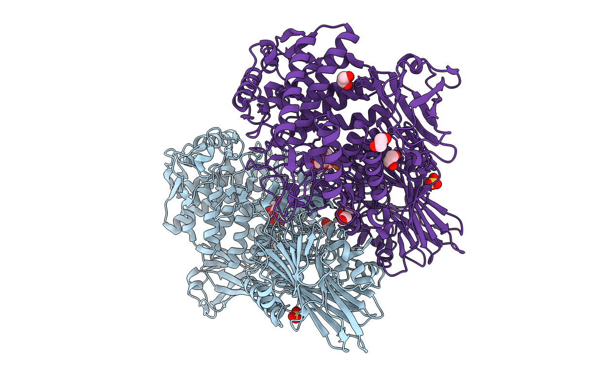
Paenibacillus sp. YM1 laminaribiose phosphorylase with alpha-glc-1-phosphate bound
Biological Source:
Source Organism:
PDB Version:
Deposition Date:
2018-05-04
Release Date:
2018-06-13
Method Details:
Experimental Method:
Resolution:
2.50 Å
R-Value Free:
0.21
R-Value Work:
0.17
R-Value Observed:
0.18
Space Group:
P 41 21 2
Macromolecular Entities
Polymer Type:polypeptide(L)
Description:Laminaribiose phosphorylase
Chain IDs:A, B
Chain Length:913
Number of Molecules:2
Biological Source:Paenibacillus sp. YM1
Primary Citation
Unravelling the Specificity of Laminaribiose Phosphorylase from Paenibacillus sp. YM-1 towards Donor Substrates Glucose/Mannose 1-Phosphate by Using X-ray Crystallography and Saturation Transfer Difference NMR Spectroscopy.
Chembiochem 20 181 192 (2019)
PMID: 29856496 DOI: 10.1002/cbic.201800260

Abstact

Glycoside phosphorylases (GPs) carry out a reversible phosphorolysis of carbohydrates into oligosaccharide acceptors and the corresponding sugar 1-phosphates. The reversibility of the reaction enables the use of GPs as biocatalysts for carbohydrate synthesis. Glycosyl hydrolase family 94 (GH94), which only comprises GPs, is one of the most studied GP families that have been used as biocatalysts for carbohydrate synthesis, in academic research and in industrial production. Understanding the mechanism of GH94 enzymes is a crucial step towards enzyme engineering to improve and expand the applications of these enzymes in synthesis. In this work with a GH94 laminaribiose phosphorylase from Paenibacillus sp. YM-1 (PsLBP), we have demonstrated an enzymatic synthesis of disaccharide 1 (β-d-mannopyranosyl-(1→3)-d-glucopyranose) by using a natural acceptor glucose and noncognate donor substrate α-mannose 1-phosphate (Man1P). To investigate how the enzyme recognises different sugar 1-phosphates, the X-ray crystal structures of PsLBP in complex with Glc1P and Man1P have been solved, providing the first molecular detail of the recognition of a noncognate donor substrate by GPs, which revealed the importance of hydrogen bonding between the active site residues and hydroxy groups at C2, C4, and C6 of sugar 1-phosphates. Furthermore, we used saturation transfer difference NMR spectroscopy to support crystallographic studies on the sugar 1-phosphates, as well as to provide further insights into the PsLBP recognition of the acceptors and disaccharide products.

Legend

Protein

Chemical

Disease

Primary Citation of related structures