Deposition Date
2018-05-03
Release Date
2018-08-08
Last Version Date
2024-10-16
Entry Detail
PDB ID:
6GGM
Keywords:
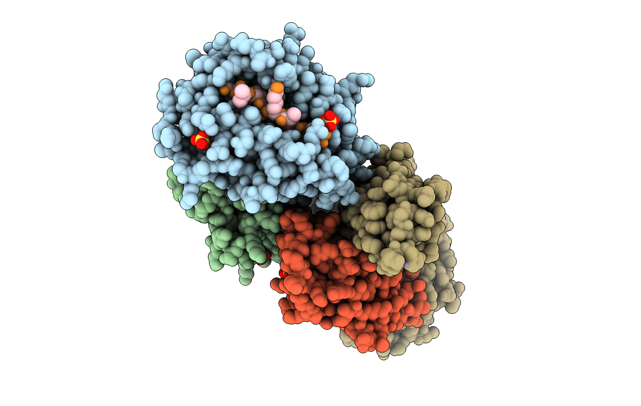
Title:
HLA-E*01:03 in complex with the Mtb44 peptide variant: Mtb44*P2-Phe.
Biological Source:
Source Organism(s):
Homo sapiens (Taxon ID: 9606)
Mycobacteriaceae (Taxon ID: 1762)
Mycobacteriaceae (Taxon ID: 1762)
Expression System(s):
Method Details:
Experimental Method:
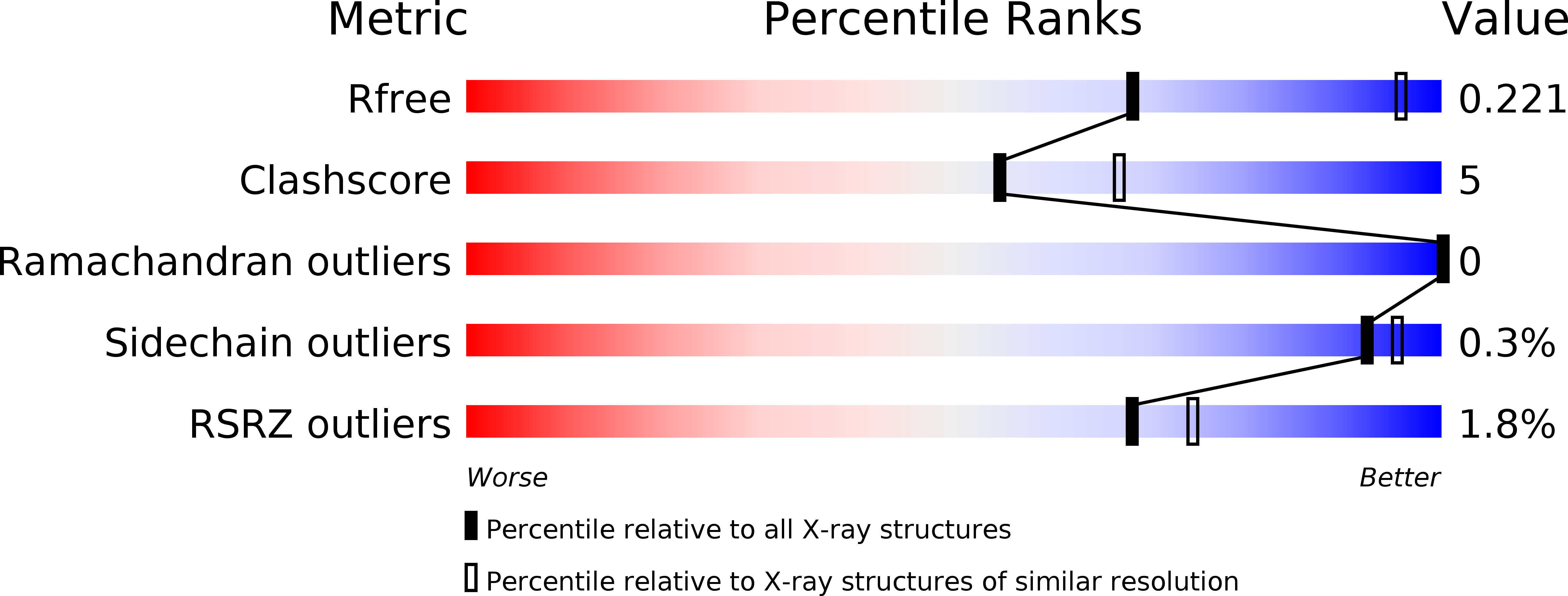
Resolution:
2.73 Å
R-Value Free:
0.22
R-Value Work:
0.18
R-Value Observed:
0.18
Space Group:
P 1