6GG9 image
Deposition Date 2018-05-03
Release Date 2018-07-25
Last Version Date 2024-01-17
Entry Detail
PDB ID:
6GG9
Title:
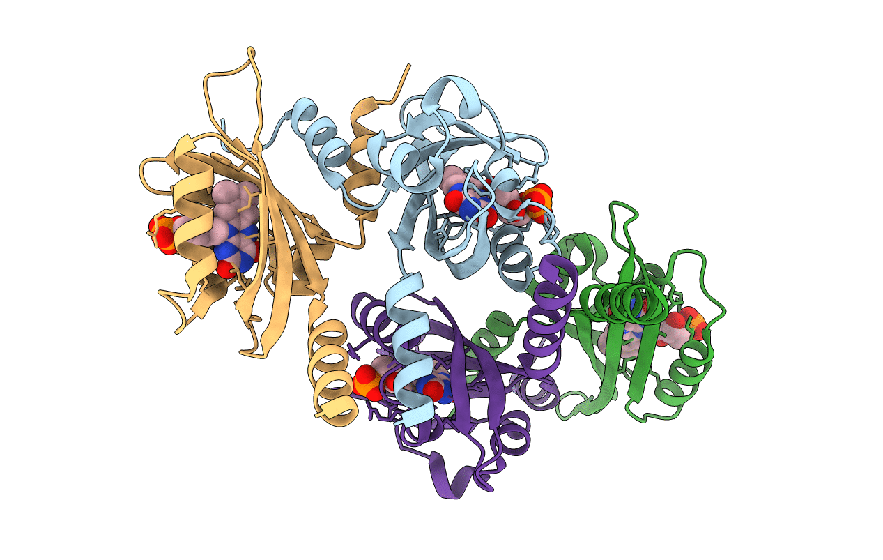
Crystal structures of a short blue light photoreceptor protein PpSB1-LOV mutant (dark state) - R61H/R66I
Biological Source:
Source Organism(s):
Method Details:
Experimental Method:
Resolution:
2.04 Å
R-Value Free:
0.23
R-Value Work:
0.19
R-Value Observed:
0.19
Space Group:
C 1 2 1
Macromolecular Entities
Structures with similar UniProt ID
Protein Blast
Polymer Type:polypeptide(L)
Molecule:Sensory box protein
Gene (Uniprot):PP_4629
Mutagens:R61H, R66I
Chain IDs:A, B, C, D
Chain Length:162
Number of Molecules:4
Biological Source:Pseudomonas putida
Ligand Molecules
Primary Citation
Small-angle X-ray scattering study of the kinetics of light-dark transition in a LOV protein.
PLoS ONE 13 e0200746 e0200746 (2018)
PMID: 30011332 DOI: 10.1371/journal.pone.0200746

Abstact

Light, oxygen, voltage (LOV) photoreceptors consist of conserved photo-responsive domains in bacteria, archaea, plants and fungi, and detect blue-light via a flavin cofactor. We investigated the blue-light induced conformational transition of the dimeric photoreceptor PpSB1-LOV-R66I from Pseudomonas putida in solution by using small-angle X-ray scattering (SAXS). SAXS experiments of the fully populated light- and dark-states under steady-state conditions revealed significant structural differences between the two states that are in agreement with the known structures determined by crystallography. We followed the transition from the light- to the dark-state by using SAXS measurements in real-time. A two-state model based on the light- and dark-state conformations could describe the measured time-course SAXS data with a relaxation time τREC of ~ 34 to 35 min being larger than the recovery time found with UV/vis spectroscopy. Unlike the flavin chromophore-based UV/vis method that is sensitive to the local chromophore environment in flavoproteins, SAXS-based assay depends on protein conformational changes and provides with an alternative to measure the recovery kinetics.

Legend

Protein

Chemical

Disease

Primary Citation of related structures
Feedback Form
Name
Email
Institute
Feedback