Deposition Date
2018-04-27
Release Date
2019-08-21
Last Version Date
2024-10-16
Entry Detail
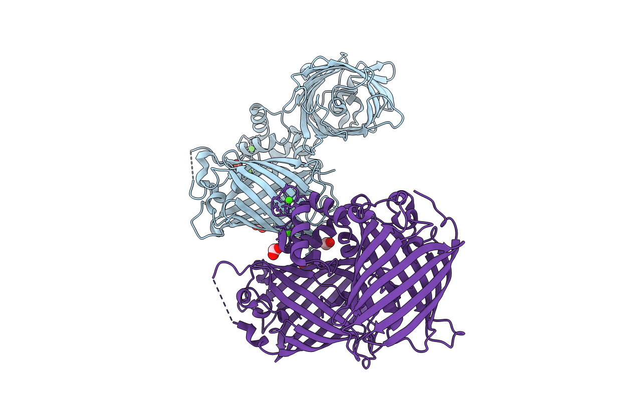
Biological Source:
Source Organism(s):
Aequorea victoria (Taxon ID: 6100)
Opsanus tau (Taxon ID: 8068)
Opsanus tau (Taxon ID: 8068)
Expression System(s):
Method Details:
Experimental Method:
Resolution:
2.47 Å
R-Value Free:
0.23
R-Value Work:
0.20
R-Value Observed:
0.20
Space Group:
P 21 21 21