6GDG image
Deposition Date 2018-04-23
Release Date 2018-05-16
Last Version Date 2024-11-20
Entry Detail
PDB ID:
6GDG
Title:
Cryo-EM structure of the adenosine A2A receptor bound to a miniGs heterotrimer
Biological Source:
Source Organism:
Escherichia coli (Taxon ID: 562)
Homo sapiens (Taxon ID: 9606)
Lama glama (Taxon ID: 9844)
Host Organism:
Method Details:
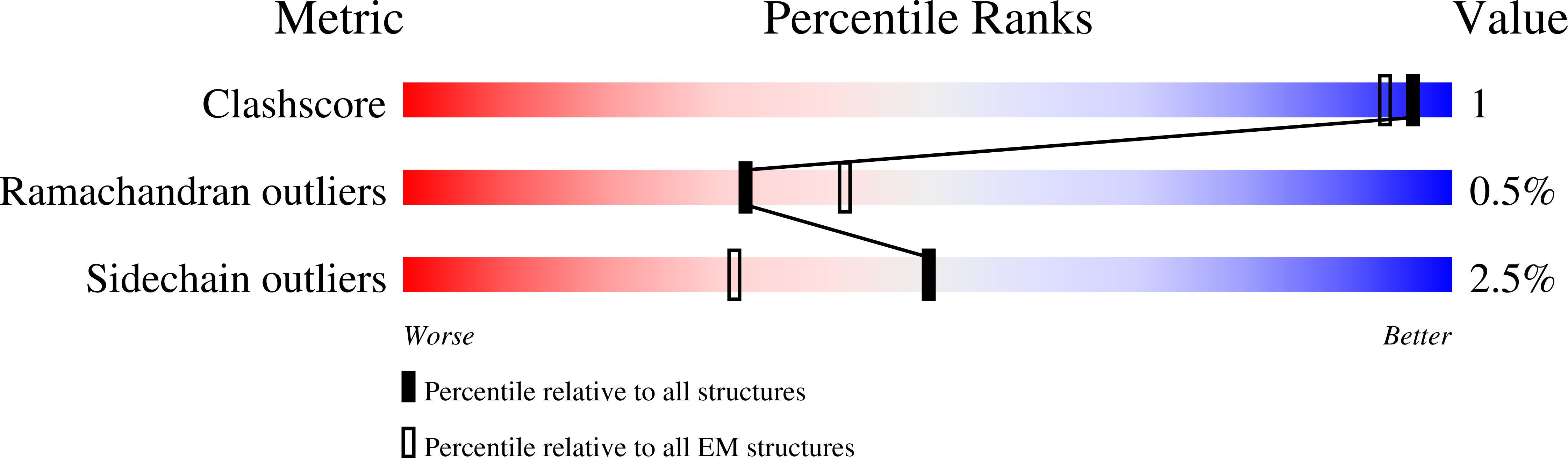
Experimental Method:
Resolution:
4.11 Å
Aggregation State:
PARTICLE
Reconstruction Method:
SINGLE PARTICLE
Macromolecular Entities
Polymer Type:polypeptide(L)
Molecule:TrxA,Adenosine receptor A2a
Gene (Uniprot):ADORA2A
Chain IDs:A
Chain Length:479
Number of Molecules:1
Biological Source:Escherichia coli, Homo sapiens
Polymer Type:polypeptide(L)
Molecule:Guanine nucleotide-binding protein G(I)/G(S)/G(T) subunit beta-1
Gene (Uniprot):GNB1
Chain IDs:B
Chain Length:339
Number of Molecules:1
Biological Source:Homo sapiens
Polymer Type:polypeptide(L)
Molecule:Guanine nucleotide-binding protein G(I)/G(S)/G(O) subunit gamma-2
Gene (Uniprot):GNG2
Chain IDs:C
Chain Length:71
Number of Molecules:1
Biological Source:Homo sapiens
Polymer Type:polypeptide(L)
Molecule:Guanine nucleotide-binding protein G(s) subunit alpha isoforms short,Guanine nucleotide-binding protein G(s) subunit alpha isoforms short
Gene (Uniprot):GNAS
Chain IDs:D
Chain Length:248
Number of Molecules:1
Biological Source:Homo sapiens
Polymer Type:polypeptide(L)
Molecule:nanobody Nb35
Chain IDs:E
Chain Length:156
Number of Molecules:1
Biological Source:Lama glama
Ligand Molecules
Primary Citation
Cryo-EM structure of the adenosine A2Areceptor coupled to an engineered heterotrimeric G protein.
Elife 7 ? ? (2018)
PMID: 29726815 DOI: 10.7554/eLife.35946

Abstact

The adenosine A2A receptor (A2AR) is a prototypical G protein-coupled receptor (GPCR) that couples to the heterotrimeric G protein GS. Here, we determine the structure by electron cryo-microscopy (cryo-EM) of A2AR at pH 7.5 bound to the small molecule agonist NECA and coupled to an engineered heterotrimeric G protein, which contains mini-GS, the βγ subunits and nanobody Nb35. Most regions of the complex have a resolution of ~3.8 Å or better. Comparison with the 3.4 Å resolution crystal structure shows that the receptor and mini-GS are virtually identical and that the density of the side chains and ligand are of comparable quality. However, the cryo-EM density map also indicates regions that are flexible in comparison to the crystal structures, which unexpectedly includes regions in the ligand binding pocket. In addition, an interaction between intracellular loop 1 of the receptor and the β subunit of the G protein was observed.

Legend

Protein

Chemical

Disease

Primary Citation of related structures