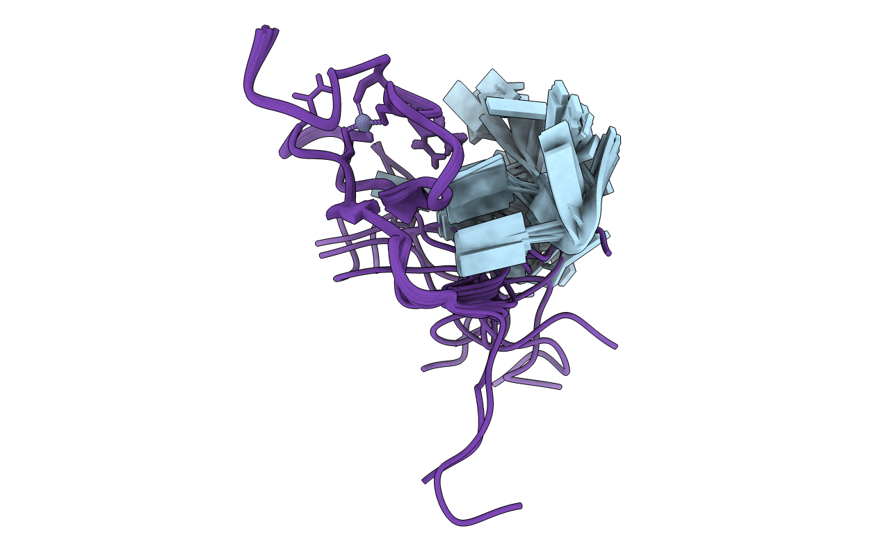
6G99 image
Entry Detail
PDB ID:
6G99
Title:
Solution structure of FUS-ZnF bound to UGGUG
Biological Source:
Source Organism:
Host Organism:
PDB Version:
Deposition Date:
2018-04-10
Release Date:
2019-02-20
Method Details:
Experimental Method:
Conformers Calculated:
500
Conformers Submitted:
20
Selection Criteria:
structures with the lowest energy
Macromolecular Entities
Polymer Type:polyribonucleotide
Description:RNA (5'-R(*UP*GP*GP*UP*G)-3')
Chain IDs:B (auth: A)
Chain Length:5
Number of Molecules:1
Biological Source:Homo sapiens
Polymer Type:polypeptide(L)
Description:RNA-binding protein FUS
Chain IDs:A (auth: B)
Chain Length:41
Number of Molecules:1
Biological Source:Homo sapiens
Ligand Molecules
Primary Citation
The Solution Structure of FUS Bound to RNA Reveals a Bipartite Mode of RNA Recognition with Both Sequence and Shape Specificity.
Mol. Cell 73 490 504.e6 (2019)
PMID: 30581145 DOI: 10.1016/j.molcel.2018.11.012

Abstact

Fused in sarcoma (FUS) is an RNA binding protein involved in regulating many aspects of RNA processing and linked to several neurodegenerative diseases. Transcriptomics studies indicate that FUS binds a large variety of RNA motifs, suggesting that FUS RNA binding might be quite complex. Here, we present solution structures of FUS zinc finger (ZnF) and RNA recognition motif (RRM) domains bound to RNA. These structures show a bipartite binding mode of FUS comprising of sequence-specific recognition of a NGGU motif via the ZnF and an unusual shape recognition of a stem-loop RNA via the RRM. In addition, sequence-independent interactions via the RGG repeats significantly increase binding affinity and promote destabilization of structured RNA conformation, enabling additional binding. We further show that disruption of the RRM and ZnF domains abolishes FUS function in splicing. Altogether, our results rationalize why deciphering the RNA binding mode of FUS has been so challenging.

Legend

Protein

Chemical

Disease

Primary Citation of related structures