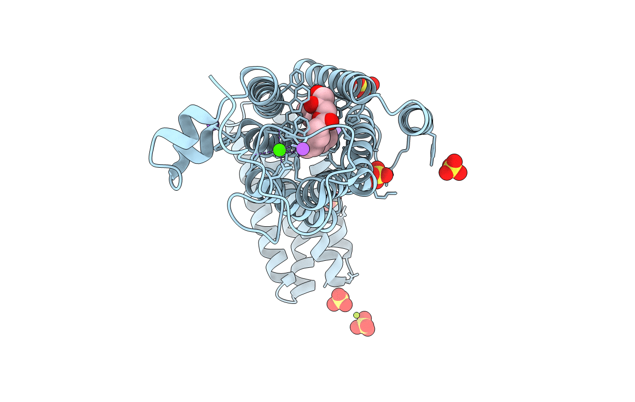
6G7O image
Deposition Date 2018-04-06
Release Date 2019-01-02
Last Version Date 2024-11-06
Entry Detail
PDB ID:
6G7O
Title:
Crystal structure of human alkaline ceramidase 3 (ACER3) at 2.7 Angstrom resolution
Biological Source:
Source Organism:
Homo sapiens (Taxon ID: 9606)
Escherichia coli (Taxon ID: 562)
Host Organism:
Method Details:
Experimental Method:
Resolution:
2.70 Å
R-Value Free:
0.27
R-Value Work:
0.24
R-Value Observed:
0.24
Space Group:
C 2 2 21
Macromolecular Entities
Polymer Type:polypeptide(L)
Molecule:Alkaline ceramidase 3,Soluble cytochrome b562
Gene (Uniprot):cybC, ACER3
Chain IDs:A
Chain Length:350
Number of Molecules:1
Biological Source:Homo sapiens, Escherichia coli
Primary Citation
Structure of a human intramembrane ceramidase explains enzymatic dysfunction found in leukodystrophy.
Nat Commun 9 5437 5437 (2018)
PMID: 30575723 DOI: 10.1038/s41467-018-07864-w

Abstact

Alkaline ceramidases (ACERs) are a class of poorly understood transmembrane enzymes controlling the homeostasis of ceramides. They are implicated in human pathophysiology, including progressive leukodystrophy, colon cancer as well as acute myeloid leukemia. We report here the crystal structure of the human ACER type 3 (ACER3). Together with computational studies, the structure reveals that ACER3 is an intramembrane enzyme with a seven transmembrane domain architecture and a catalytic Zn2+ binding site in its core, similar to adiponectin receptors. Interestingly, we uncover a Ca2+ binding site physically and functionally connected to the Zn2+ providing a structural explanation for the known regulatory role of Ca2+ on ACER3 enzymatic activity and for the loss of function in E33G-ACER3 mutant found in leukodystrophic patients.

Legend

Protein

Chemical

Disease

Primary Citation of related structures