6G7J image
Deposition Date 2018-04-06
Release Date 2018-06-27
Last Version Date 2024-10-23
Entry Detail
PDB ID:
6G7J
Keywords:
Title:
Retinal isomerization in bacteriorhodopsin revealed by a femtosecond X-ray laser: 457-646 fs state structure
Biological Source:
Method Details:
Experimental Method:
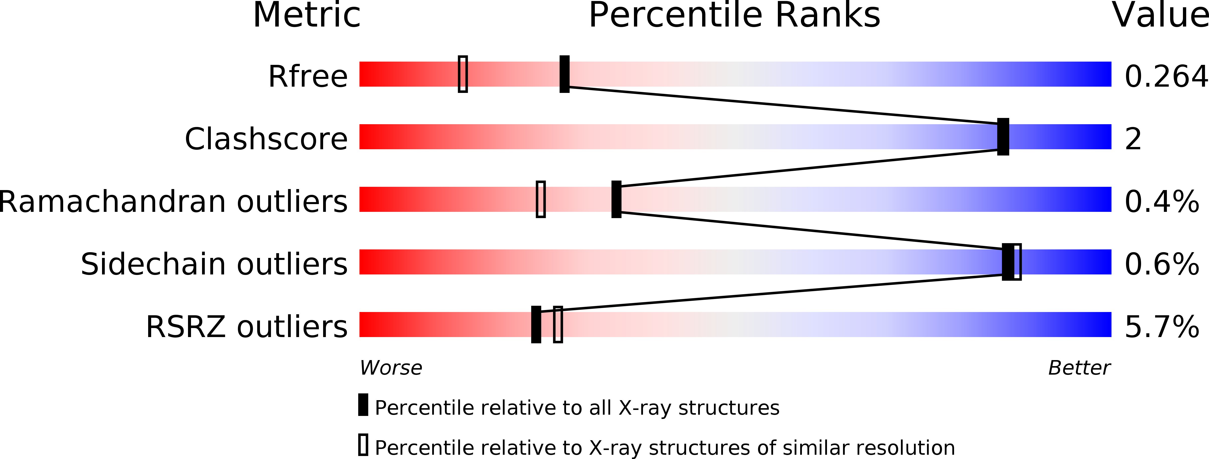
Resolution:
1.90 Å
R-Value Free:
0.26
R-Value Work:
0.20
R-Value Observed:
0.20
Space Group:
P 63
Macromolecular Entities
Structures with similar UniProt ID
Protein Blast
Polymer Type:polypeptide(L)
Molecule:Bacteriorhodopsin
Gene (Uniprot):bop
Chain IDs:A
Chain Length:262
Number of Molecules:1
Biological Source:Halobacterium salinarum (strain ATCC 700922 / JCM 11081 / NRC-1)
Primary Citation

Abstact

Ultrafast isomerization of retinal is the primary step in photoresponsive biological functions including vision in humans and ion transport across bacterial membranes. We used an x-ray laser to study the subpicosecond structural dynamics of retinal isomerization in the light-driven proton pump bacteriorhodopsin. A series of structural snapshots with near-atomic spatial resolution and temporal resolution in the femtosecond regime show how the excited all-trans retinal samples conformational states within the protein binding pocket before passing through a twisted geometry and emerging in the 13-cis conformation. Our findings suggest ultrafast collective motions of aspartic acid residues and functional water molecules in the proximity of the retinal Schiff base as a key facet of this stereoselective and efficient photochemical reaction.

Legend

Protein

Chemical

Disease

Primary Citation of related structures