Deposition Date
2018-03-27
Release Date
2019-01-30
Last Version Date
2024-05-08
Entry Detail
PDB ID:
6G4J
Keywords:
Title:
Structure of the protein kinase YabT from Bacillus subtilis in complex with an alphaREP crystallization helper
Biological Source:
Source Organism(s):
Bacillus subtilis (strain 168) (Taxon ID: 224308)
synthetic construct (Taxon ID: 32630)
synthetic construct (Taxon ID: 32630)
Expression System(s):
Method Details:
Experimental Method:
Resolution:
1.60 Å
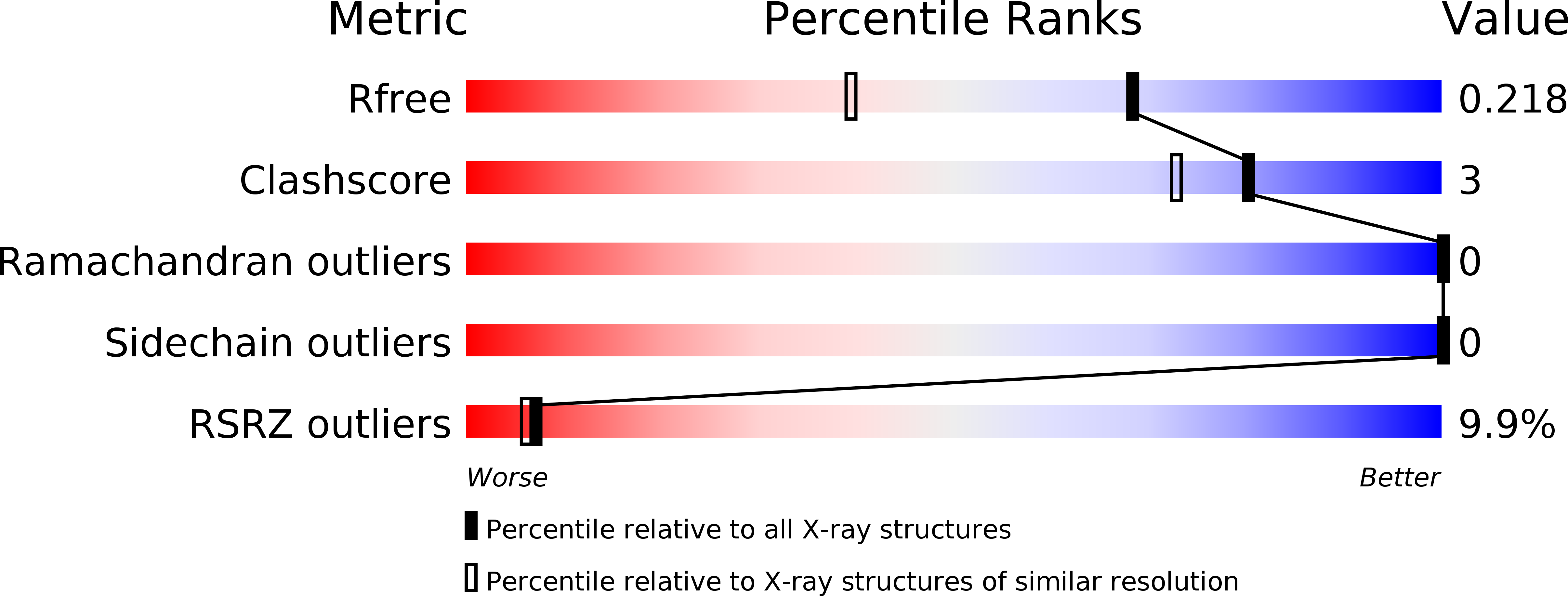
R-Value Free:
0.21
R-Value Work:
0.18
R-Value Observed:
0.19
Space Group:
P 21 21 2