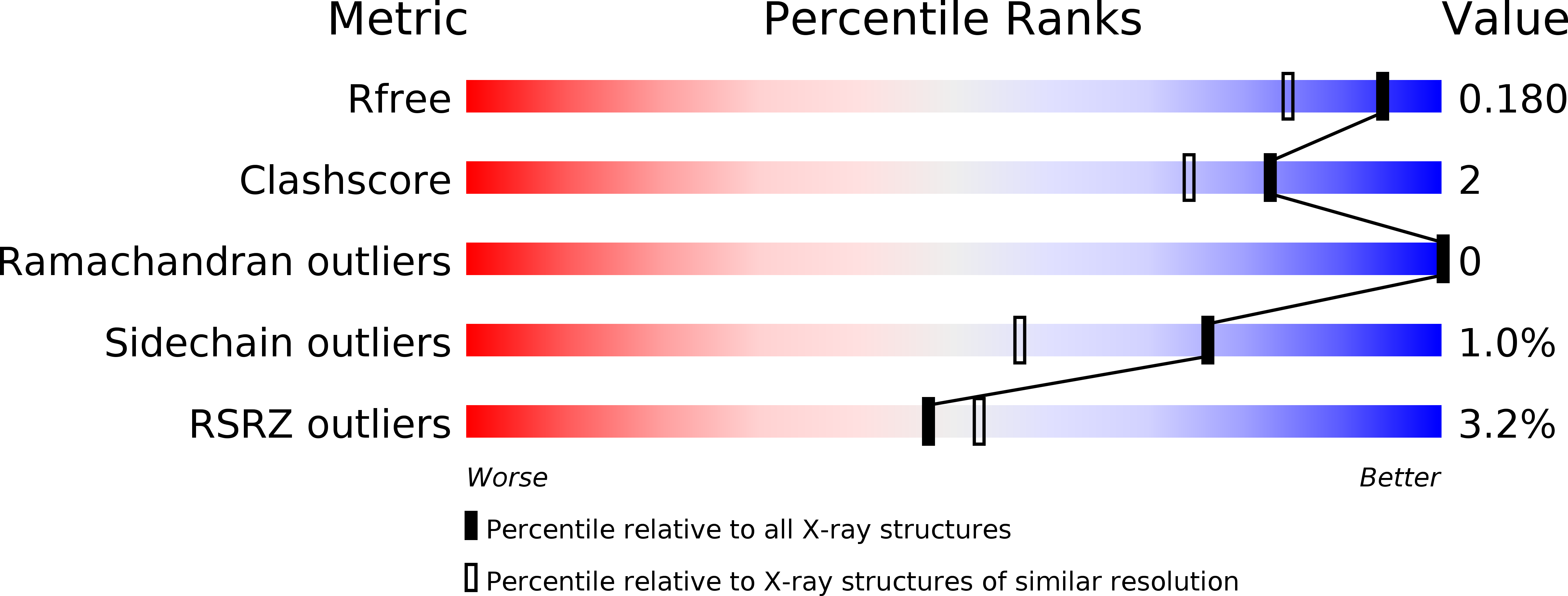
6G47 image
Deposition Date 2018-03-26
Release Date 2018-05-02
Last Version Date 2024-01-17
Entry Detail
PDB ID:
6G47
Keywords:
Title:
Crystal Structure of Human Adenovirus 52 Short Fiber Knob in Complex with alpha-(2,8)-Trisialic Acid (DP3)
Biological Source:
Source Organism:
Host Organism:
Method Details:
Experimental Method:
Resolution:
1.50 Å
R-Value Free:
0.17
R-Value Work:
0.16
R-Value Observed:
0.16
Space Group:
P 21 21 21
Macromolecular Entities
Structures with similar UniProt ID
Protein Blast
Polymer Type:polypeptide(L)
Molecule:Fiber-1
Mutagens:N-terminal His-tag, partially resolved
Chain IDs:A, B, C
Chain Length:209
Number of Molecules:3
Biological Source:Human adenovirus 52
Primary Citation
Polysialic acid is a cellular receptor for human adenovirus 52.
Proc. Natl. Acad. Sci. U.S.A. 115 E4264 E4273 (2018)
PMID: 29674446 DOI: 10.1073/pnas.1716900115

Abstact

Human adenovirus 52 (HAdV-52) is one of only three known HAdVs equipped with both a long and a short fiber protein. While the long fiber binds to the coxsackie and adenovirus receptor, the function of the short fiber in the virus life cycle is poorly understood. Here, we show, by glycan microarray analysis and cellular studies, that the short fiber knob (SFK) of HAdV-52 recognizes long chains of α-2,8-linked polysialic acid (polySia), a large posttranslational modification of selected carrier proteins, and that HAdV-52 can use polySia as a receptor on target cells. X-ray crystallography, NMR, molecular dynamics simulation, and structure-guided mutagenesis of the SFK reveal that the nonreducing, terminal sialic acid of polySia engages the protein with direct contacts, and that specificity for polySia is achieved through subtle, transient electrostatic interactions with additional sialic acid residues. In this study, we present a previously unrecognized role for polySia as a cellular receptor for a human viral pathogen. Our detailed analysis of the determinants of specificity for this interaction has general implications for protein-carbohydrate interactions, particularly concerning highly charged glycan structures, and provides interesting dimensions on the biology and evolution of members of Human mastadenovirus G.

Legend

Protein

Chemical

Disease

Primary Citation of related structures