6G03 image
Entry Detail
PDB ID:
6G03
Title:
NMR Solution Structure of yeast TSR2(1-152)
Biological Source:
Source Organism:
PDB Version:
Deposition Date:
2018-03-16
Release Date:
2018-09-19
Method Details:
Experimental Method:
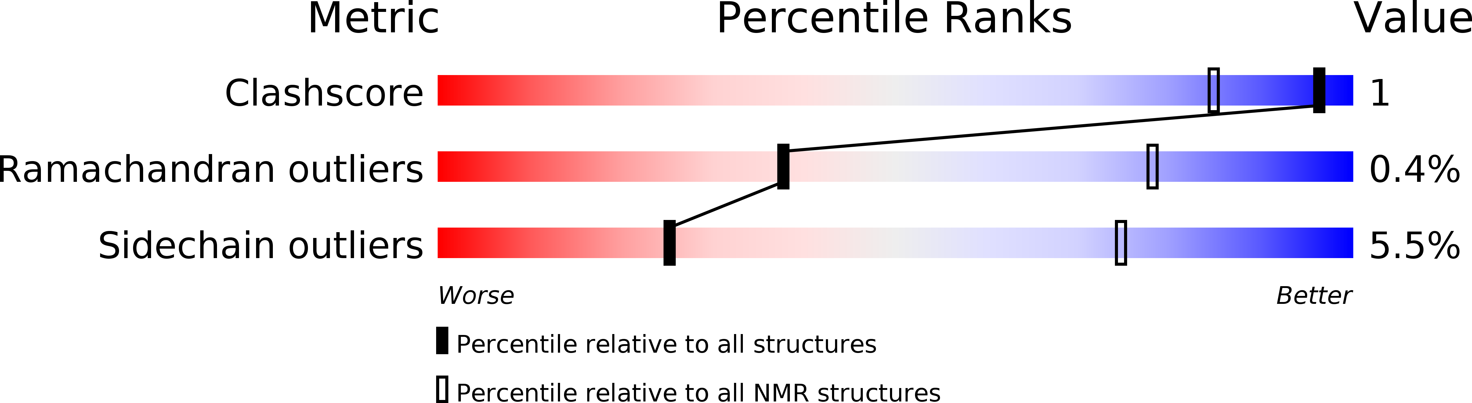
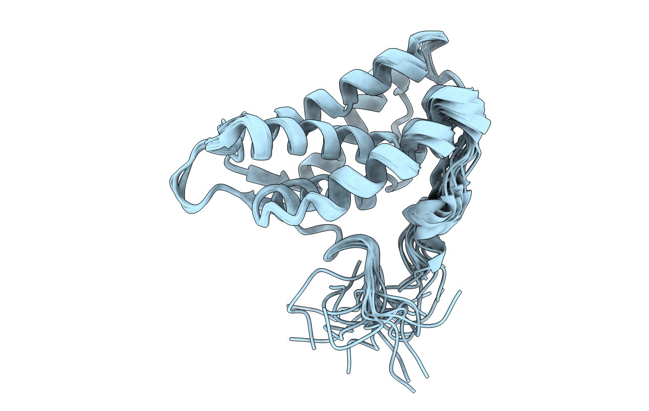
Conformers Calculated:
100
Conformers Submitted:
20
Selection Criteria:
structures with the lowest energy
Macromolecular Entities
Polymer Type:polypeptide(L)
Description:Pre-rRNA-processing protein TSR2
Chain IDs:A
Chain Length:156
Number of Molecules:1
Biological Source:Saccharomyces cerevisiae
Ligand Molecules
Primary Citation
Molecular basis for disassembly of an importin:ribosomal protein complex by the escortin Tsr2.
Nat Commun 9 3669 3669 (2018)
PMID: 30201955 DOI: 10.1038/s41467-018-06160-x

Abstact

Disordered extensions at the termini and short internal insertions distinguish eukaryotic ribosomal proteins (r-proteins) from their anucleated archaeal counterparts. Here, we report an NMR structure of such a eukaryotic-specific segment (ESS) in the r-protein eS26 in complex with the escortin Tsr2. The structure reveals how ESS attracts Tsr2 specifically to importin:eS26 complexes entering the nucleus in order to trigger non-canonical RanGTP-independent disassembly. Tsr2 then sequesters the released eS26 and prevents rebinding to the importin, providing an alternative allosteric mechanism to terminate the process of nuclear import. Notably, a Diamond-Blackfan anemia-associated Tsr2 mutant protein is impaired in binding to ESS, unveiling a critical role for this interaction in human hematopoiesis. We propose that eS26-ESS and Tsr2 are components of a nuclear sorting system that co-evolved with the emergence of the nucleocytoplasmic barrier and transport carriers.

Legend

Protein

Chemical

Disease

Primary Citation of related structures