6FZV image
Entry Detail
PDB ID:
6FZV
Title:
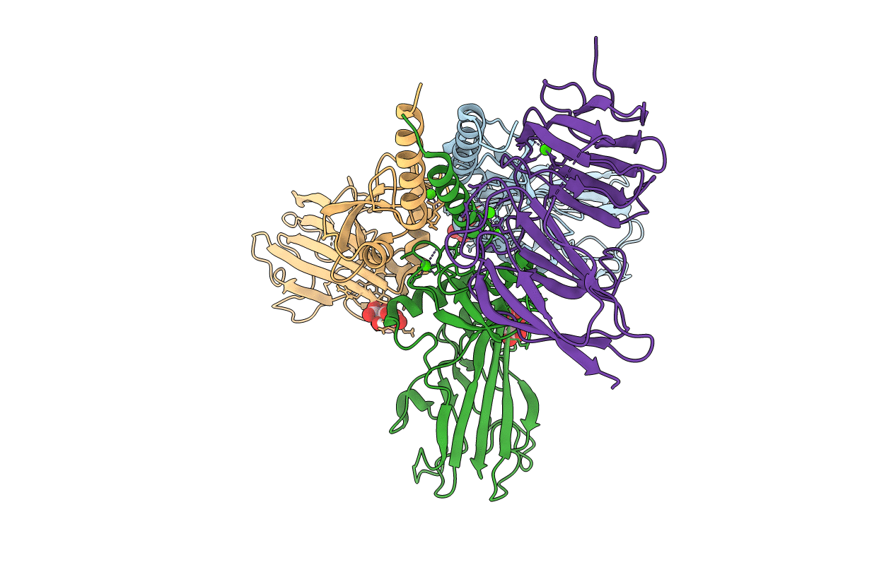
Crystal structure of the metalloproteinase enhancer PCPE-1 bound to the procollagen C propeptide trimer (short)
Biological Source:
Source Organism:
Host Organism:
PDB Version:
Deposition Date:
2018-03-15
Release Date:
2018-07-04
Method Details:
Experimental Method:
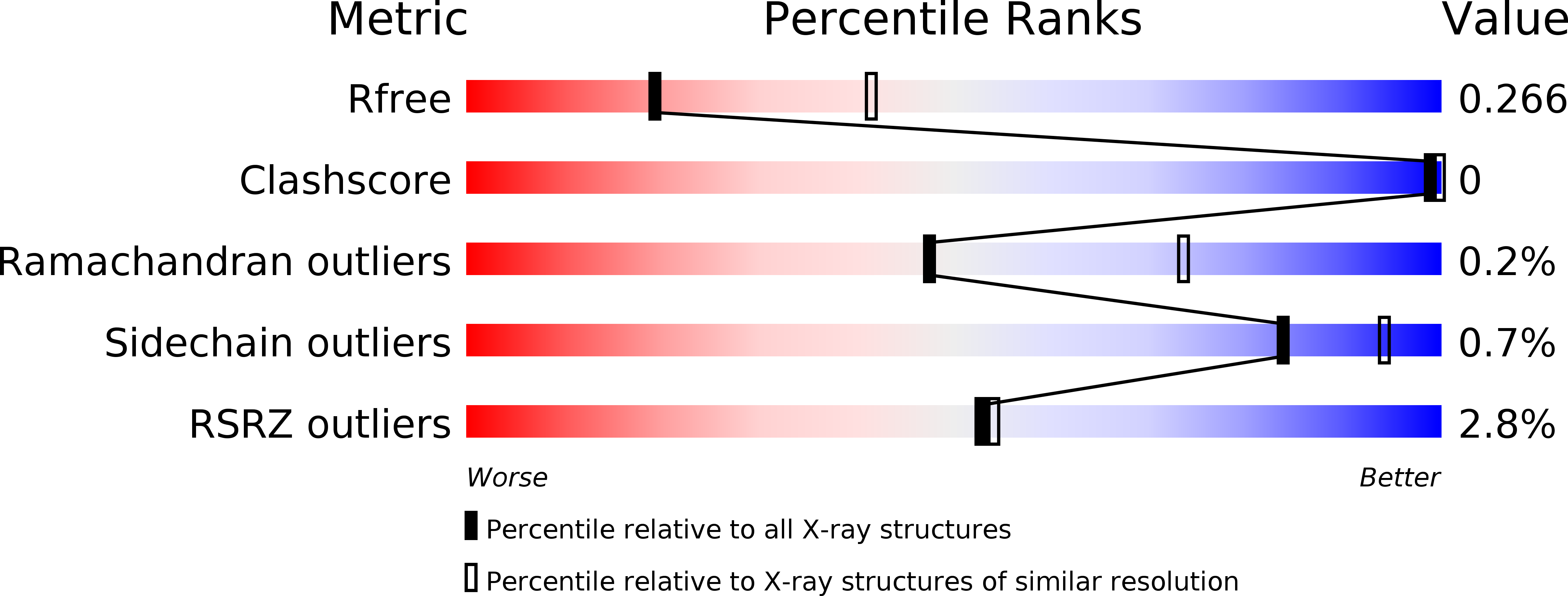
Resolution:
2.70 Å
R-Value Free:
0.25
R-Value Work:
0.22
R-Value Observed:
0.22
Space Group:
P 21 21 21
Macromolecular Entities
Polymer Type:polypeptide(L)
Description:Collagen alpha-1(III) chain
Chain IDs:A, B, C
Chain Length:256
Number of Molecules:3
Biological Source:Homo sapiens
Polymer Type:polypeptide(L)
Description:Procollagen C-endopeptidase enhancer 1
Chain IDs:D
Chain Length:265
Number of Molecules:1
Biological Source:Homo sapiens
Primary Citation
Structural Basis for the Acceleration of Procollagen Processing by Procollagen C-Proteinase Enhancer-1.
Structure 26 1384 1392.e3 (2018)
PMID: 30078642 DOI: 10.1016/j.str.2018.06.011

Abstact

Procollagen C-proteinase enhancer-1 (PCPE-1) is a secreted protein that specifically accelerates proteolytic release of the C-propeptides from fibrillar procollagens, a crucial step in fibril assembly. As such, it is a potential therapeutic target to improve tissue repair and prevent fibrosis, a major cause of mortality worldwide. Here we present the crystal structure of the active CUB1CUB2 fragment of PCPE-1 bound to the C-propeptide trimer of procollagen III (CPIII). This shows that the two CUB domains bind to two different chains of CPIII and that the N-terminal region of one CPIII chain, close to the proteolytic cleavage site, lies in the cleft between CUB1 and CUB2. This suggests that enhancing activity involves unraveling of this chain from the rest of the trimer, thus facilitating the action of the proteinase involved. Support for this hypothesis comes from site-directed mutagenesis, enzyme assays, binding studies, and molecular modeling.

Legend

Protein

Chemical

Disease

Primary Citation of related structures