Deposition Date
2018-03-15
Release Date
2019-02-20
Last Version Date
2024-06-19
Entry Detail
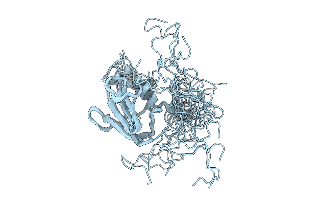
PDB ID:
6FZK
Keywords:
Title:
NMR structure of UB2H, regulatory domain of PBP1b from E. coli
Biological Source:
Source Organism(s):
Escherichia coli K-12 (Taxon ID: 83333)
Expression System(s):
Method Details:
Experimental Method:
Conformers Calculated:
750
Conformers Submitted:
20
Selection Criteria:
20