
Deposition Date
2018-03-13
Release Date
2018-07-04
Last Version Date
2024-05-08
Entry Detail
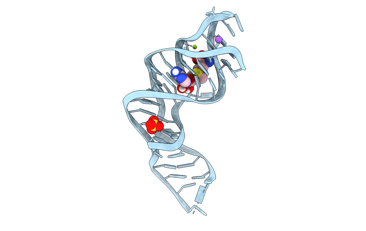
Biological Source:
Source Organism(s):
Candidatus Pelagibacter ubique HTCC1062 (Taxon ID: 335992)
Method Details:
Experimental Method:
Resolution:
2.50 Å
R-Value Free:
0.26
R-Value Work:
0.23
R-Value Observed:
0.23
Space Group:
P 41 2 2
