Deposition Date
2018-03-09
Release Date
2019-03-20
Last Version Date
2024-02-07
Entry Detail
PDB ID:
6FXU
Keywords:
Title:
Crystal structure of human transthyretin mutant T119M at pH 5.5
Biological Source:
Source Organism(s):
Homo sapiens (Taxon ID: 9606)
Expression System(s):
Method Details:
Experimental Method:
Resolution:
1.36 Å
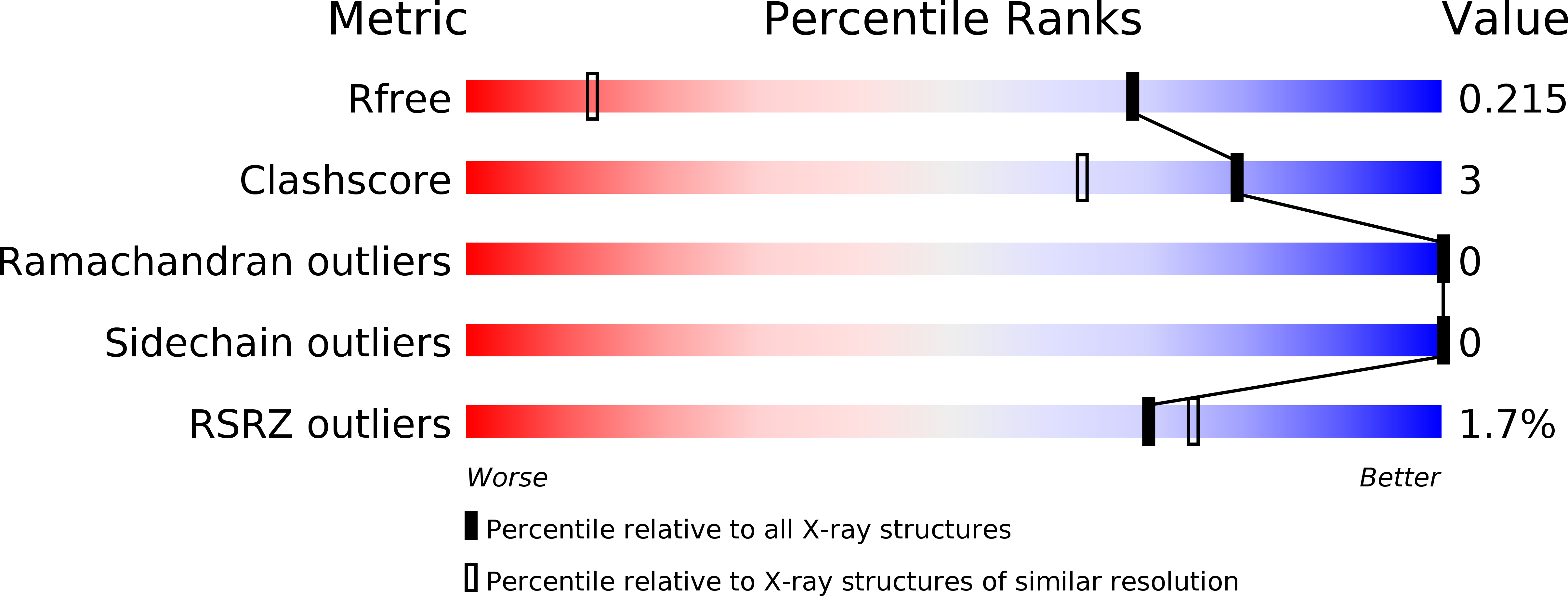
R-Value Free:
0.21
R-Value Work:
0.19
R-Value Observed:
0.19
Space Group:
P 21 21 2