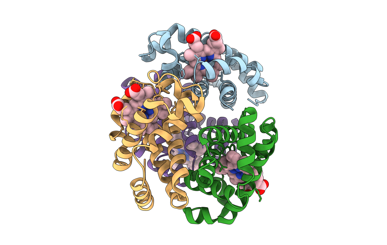
6FQF image
Entry Detail
PDB ID:
6FQF
Title:
THE X-RAY STRUCTURE OF FERRIC-BETA4 HUMAN HEMOGLOBIN
Biological Source:
Source Organism:
Host Organism:
PDB Version:
Deposition Date:
2018-02-14
Release Date:
2018-06-13
Method Details:
Experimental Method:
Resolution:
2.10 Å
R-Value Free:
0.26
R-Value Work:
0.20
R-Value Observed:
0.20
Space Group:
P 1 21 1
Macromolecular Entities
Polymer Type:polypeptide(L)
Description:Hemoglobin subunit beta
Chain IDs:A, B, C, D
Chain Length:146
Number of Molecules:4
Biological Source:Homo sapiens
Ligand Molecules
Primary Citation
Crystal structure of the ferric homotetrameric beta4human hemoglobin.
Biophys. Chem. 240 9 14 (2018)
PMID: 29857171 DOI: 10.1016/j.bpc.2018.05.003

Abstact

Spectroscopic studies carried out in the early seventies have shown that the β-homotetramer of human hemoglobin (β4-HbA) in the ferric state is a mixture of aquomet and bis-histidyl forms. Here we present the first crystal structure, solved at 2.10 Å resolution, of the oxidized form of β4-HbA. The overall quaternary structure of the protein in the ferric state is virtually indistinguishable from that of the ferrous deoxygenated and carbomonoxy forms. The structure reveals that the four hemes are exclusively in an aquomet coordination, without any trace of bis-histidyl coordination. The oxidation of β4-HbA is associated with the formation of a disulfide bridge between residues Cys112(G14) of β1/β4 and β2/β3 chains. The coordination state of β4-HbA has been compared to that known for other organisms that exhibit bis-histidyl heme coordination in the β4 state. This occurrence has been discussed in terms of different organism physiology.

Legend

Protein

Chemical

Disease

Primary Citation of related structures