Deposition Date
2018-02-07
Release Date
2018-11-07
Last Version Date
2024-05-08
Entry Detail
PDB ID:
6FOG
Keywords:
Title:
X-ray structure of homo sapiens Fumarylacetoacetate hydrolase domain containing protein 1 (FAHD1) in complex with inhibitor oxalate at 1.94A resolution.
Biological Source:
Source Organism(s):
Homo sapiens (Taxon ID: 9606)
Expression System(s):
Method Details:
Experimental Method:
Resolution:
1.94 Å
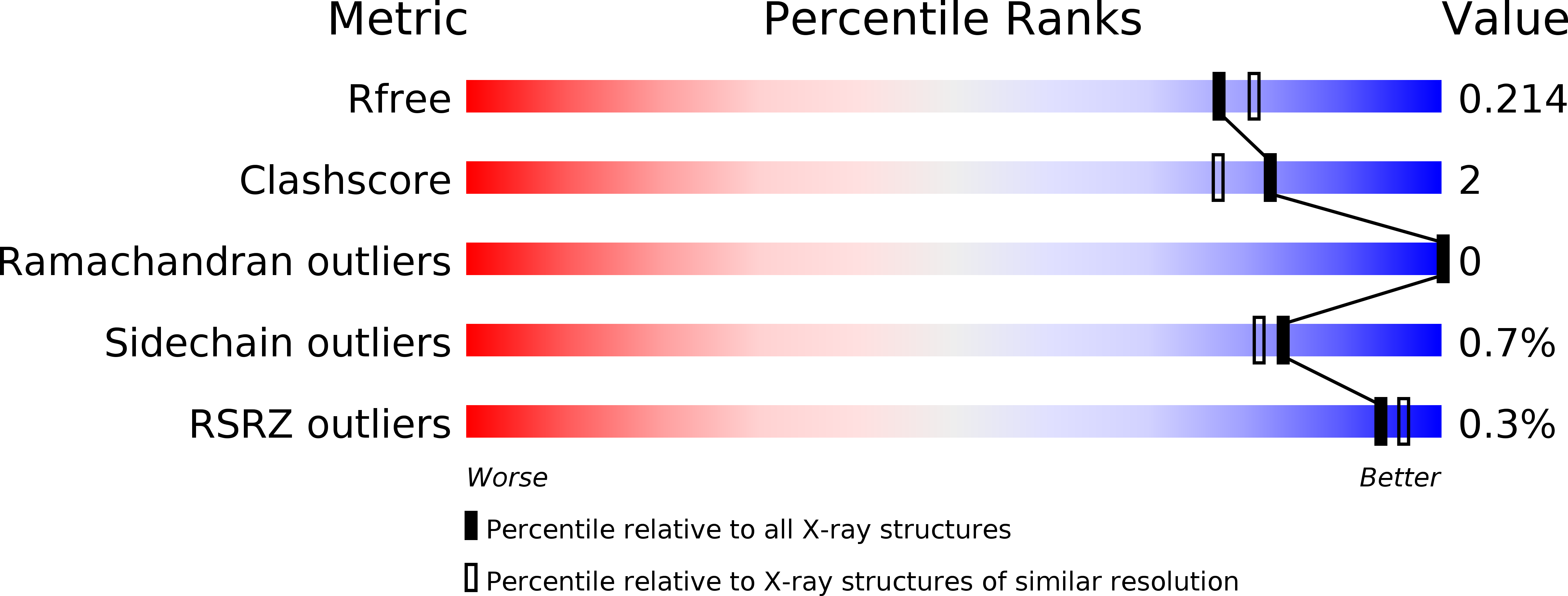
R-Value Free:
0.20
R-Value Work:
0.18
R-Value Observed:
0.18
Space Group:
P 1 21 1