Deposition Date
2018-02-06
Release Date
2019-01-23
Last Version Date
2024-01-17
Entry Detail
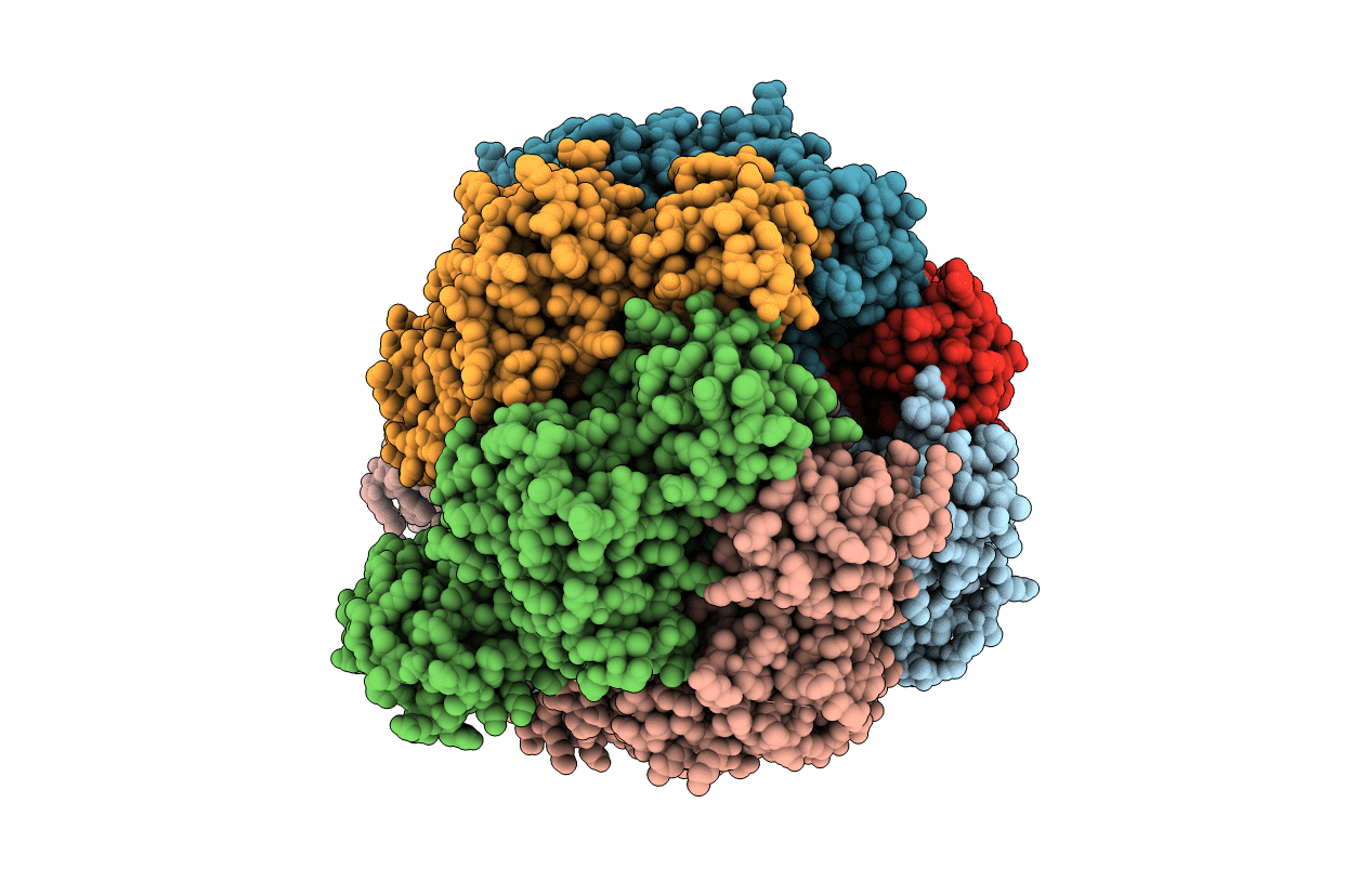
Biological Source:
Source Organism(s):
Mycolicibacterium smegmatis MC2 155 (Taxon ID: 246196)
Expression System(s):
Method Details:
Experimental Method:
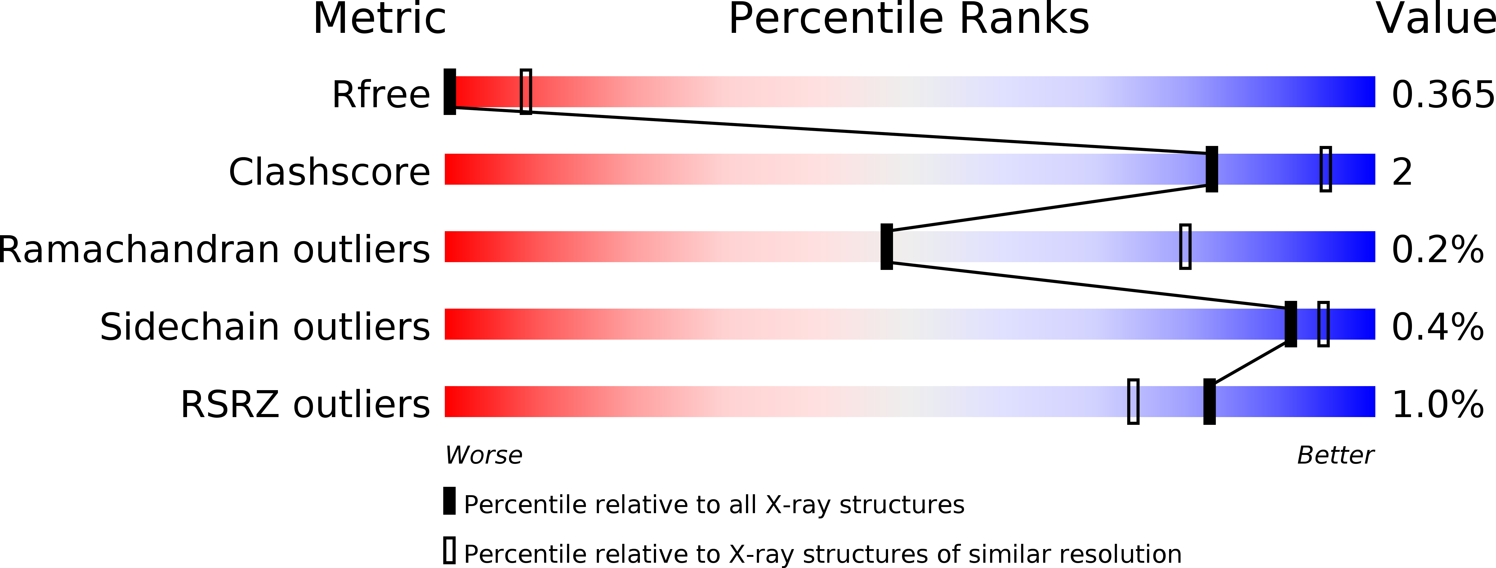
Resolution:
4.00 Å
R-Value Free:
0.36
R-Value Work:
0.33
R-Value Observed:
0.33
Space Group:
P 31 2 1