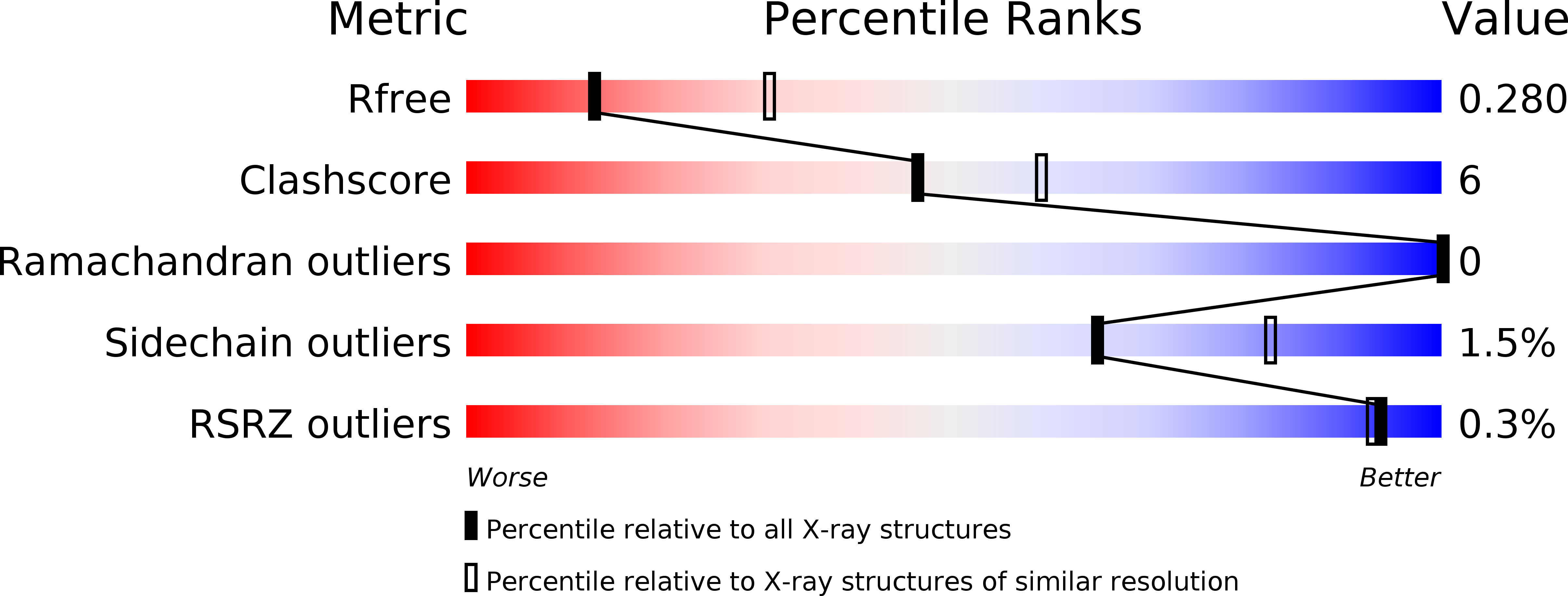
6FEL image
Deposition Date 2018-01-02
Release Date 2018-01-10
Last Version Date 2024-10-09
Entry Detail
PDB ID:
6FEL
Title:
Structure of 14-3-3 gamma in complex with CaMKK2 14-3-3 binding motif Ser511
Biological Source:
Source Organism:
Homo sapiens (Taxon ID: 9606)
Method Details:
Experimental Method:
Resolution:
2.84 Å
R-Value Free:
0.27
R-Value Work:
0.22
R-Value Observed:
0.23
Space Group:
H 3
Macromolecular Entities
Structures with similar UniProt ID
Protein Blast
Polymer Type:polypeptide(L)
Molecule:14-3-3 protein gamma
Gene (Uniprot):YWHAG
Chain IDs:A, B (auth: C), C (auth: B), D
Chain Length:236
Number of Molecules:4
Biological Source:Homo sapiens
Structures with similar UniProt ID
Protein Blast
Polymer Type:polypeptide(L)
Molecule:Calcium/calmodulin-dependent protein kinase kinase 2
Gene (Uniprot):CAMKK2
Chain IDs:E, F, G, H
Chain Length:8
Number of Molecules:4
Biological Source:Homo sapiens
Modified Residue
Compound ID Chain ID Parent Comp ID Details 2D Image
SEP E SER modified residue
Primary Citation
14-3-3 protein directly interacts with the kinase domain of calcium/calmodulin-dependent protein kinase kinase (CaMKK2).
Biochim. Biophys. Acta 1862 1612 1625 (2018)
PMID: 29649512 DOI: 10.1016/j.bbagen.2018.04.006

Abstact

BACKGROUND Calcium/calmodulin-dependent protein kinase kinase 2 (CaMKK2) is a member of the Ca2+/calmodulin-dependent kinase (CaMK) family involved in adiposity regulation, glucose homeostasis and cancer. This upstream activator of CaMKI, CaMKIV and AMP-activated protein kinase is inhibited by phosphorylation, which also triggers an association with the scaffolding protein 14-3-3. However, the role of 14-3-3 in the regulation of CaMKK2 remains unknown. METHODS The interaction between phosphorylated CaMKK2 and the 14-3-3γ protein, as well as the architecture of their complex, were studied using enzyme activity measurements, small-angle x-ray scattering (SAXS), time-resolved fluorescence spectroscopy and protein crystallography. RESULTS Our data suggest that the 14-3-3 protein binding does not inhibit the catalytic activity of phosphorylated CaMKK2 but rather slows down its dephosphorylation. Structural analysis indicated that the complex is flexible and that CaMKK2 is located outside the phosphopeptide-binding central channel of the 14-3-3γ dimer. Furthermore, 14-3-3γ appears to interact with and affect the structure of several regions of CaMKK2 outside the 14-3-3 binding motifs. In addition, the structural basis of interactions between 14-3-3 and the 14-3-3 binding motifs of CaMKK2 were elucidated by determining the crystal structures of phosphopeptides containing these motifs bound to 14-3-3. CONCLUSIONS 14-3-3γ protein directly interacts with the kinase domain of CaMKK2 and the region containing the inhibitory phosphorylation site Thr145 within the N-terminal extension. GENERAL SIGNIFICANCE Our results suggested that CaMKK isoforms differ in their 14-3-3-mediated regulations and that the interaction between 14-3-3 protein and the N-terminal 14-3-3-binding motif of CaMKK2 might be stabilized by small-molecule compounds.

Legend

Protein

Chemical

Disease

Primary Citation of related structures