6FEH image
Deposition Date 2018-01-02
Release Date 2018-02-21
Last Version Date 2024-05-15
Entry Detail
PDB ID:
6FEH
Keywords:
Title:
Solution Structure of CaM/Kv7.2-hAB Complex
Biological Source:
Source Organism:
Homo sapiens (Taxon ID: 9606)
Host Organism:
Method Details:
Experimental Method:
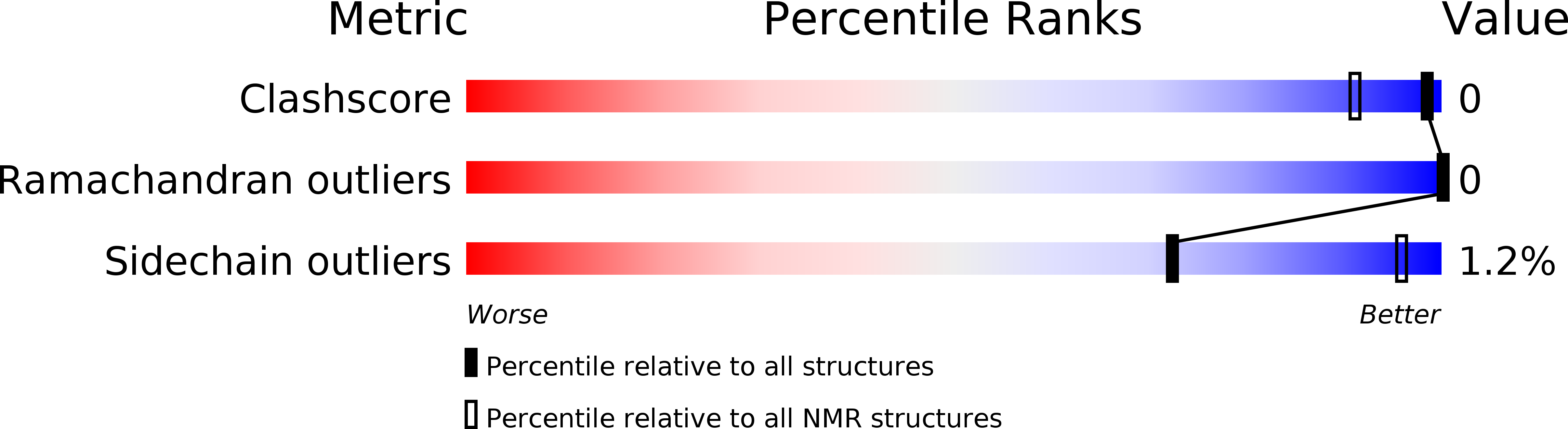
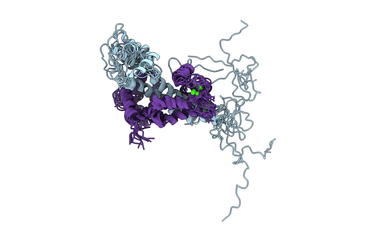
Conformers Calculated:
200
Conformers Submitted:
10
Selection Criteria:
structures with the least restraint violations
Macromolecular Entities
Structures with similar UniProt ID
Protein Blast
Polymer Type:polypeptide(L)
Molecule:Potassium voltage-gated channel subfamily KQT member 2,Potassium voltage-gated channel subfamily KQT member 2
Gene (Uniprot):KCNQ2
Mutagens:Del. R374-K493,Del. R374-K493
Chain IDs:A
Chain Length:115
Number of Molecules:1
Biological Source:Homo sapiens
Structures with similar UniProt ID
Protein Blast
Polymer Type:polypeptide(L)
Molecule:Calmodulin-1
Gene (Uniprot):CALM1
Chain IDs:B
Chain Length:149
Number of Molecules:1
Biological Source:Homo sapiens
Ligand Molecules
Primary Citation
Structural basis and energy landscape for the Ca2+gating and calmodulation of the Kv7.2 K+channel.
Proc. Natl. Acad. Sci. U.S.A. 115 2395 2400 (2018)
PMID: 29463698 DOI: 10.1073/pnas.1800235115

Abstact

The Kv7.2 (KCNQ2) channel is the principal molecular component of the slow voltage-gated, noninactivating K(+) M-current, a key controller of neuronal excitability. To investigate the calmodulin (CaM)-mediated Ca(2+) gating of the channel, we used NMR spectroscopy to structurally and dynamically describe the association of helices hA and hB of Kv7.2 with CaM, as a function of Ca(2+) concentration. The structures of the CaM/Kv7.2-hAB complex at two different calcification states are reported here. In the presence of a basal cytosolic Ca(2+) concentration (10-100 nM), only the N-lobe of CaM is Ca(2+)-loaded and the complex (representative of the open channel) exhibits collective dynamics on the millisecond time scale toward a low-populated excited state (1.5%) that corresponds to the inactive state of the channel. In response to a chemical or electrical signal, intracellular Ca(2+) levels rise up to 1-10 muM, triggering Ca(2+) association with the C-lobe. The associated conformational rearrangement is the key biological signal that shifts populations to the closed/inactive channel. This reorientation affects the C-lobe of CaM and both helices in Kv7.2, allosterically transducing the information from the Ca(2+)-binding site to the transmembrane region of the channel.

Legend

Protein

Chemical

Disease

Primary Citation of related structures