
Deposition Date
1997-09-17
Release Date
1997-11-12
Last Version Date
2024-05-22
Entry Detail
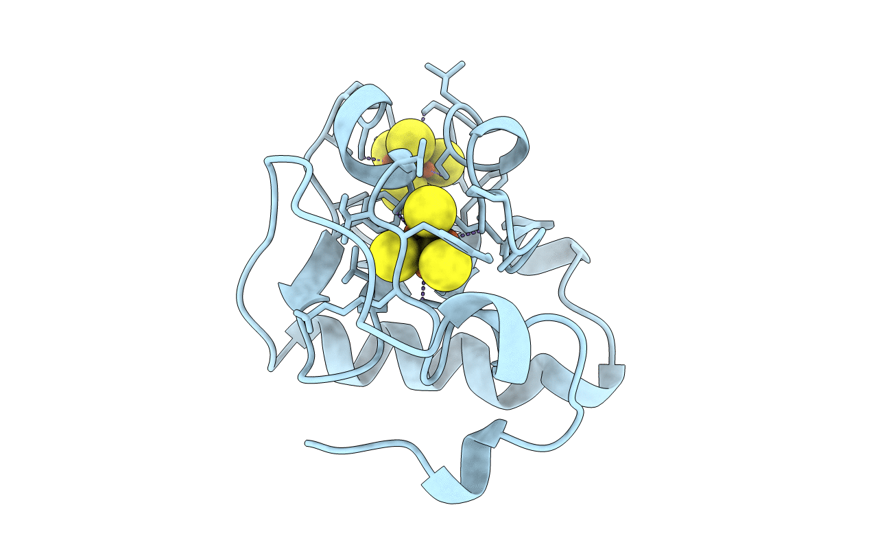
PDB ID:
6FD1
Keywords:
Title:
7-FE FERREDOXIN FROM AZOTOBACTER VINELANDII LOW TEMPERATURE, 1.35 A
Biological Source:
Source Organism(s):
Azotobacter vinelandii (Taxon ID: 354)
Method Details:
Experimental Method:
Resolution:
1.35 Å
R-Value Free:
0.21
R-Value Observed:
0.15
Space Group:
P 41 21 2
