6FBR image
Deposition Date 2017-12-19
Release Date 2018-12-05
Last Version Date 2024-11-13
Entry Detail
PDB ID:
6FBR
Keywords:
Title:
Crystal Structure of the Human Retinoid X Receptor DNA-Binding Domain Bound to the Human MEp DR1 Response Element, pH 4.2
Biological Source:
Source Organism:
Homo sapiens (Taxon ID: 9606)
Method Details:
Experimental Method:
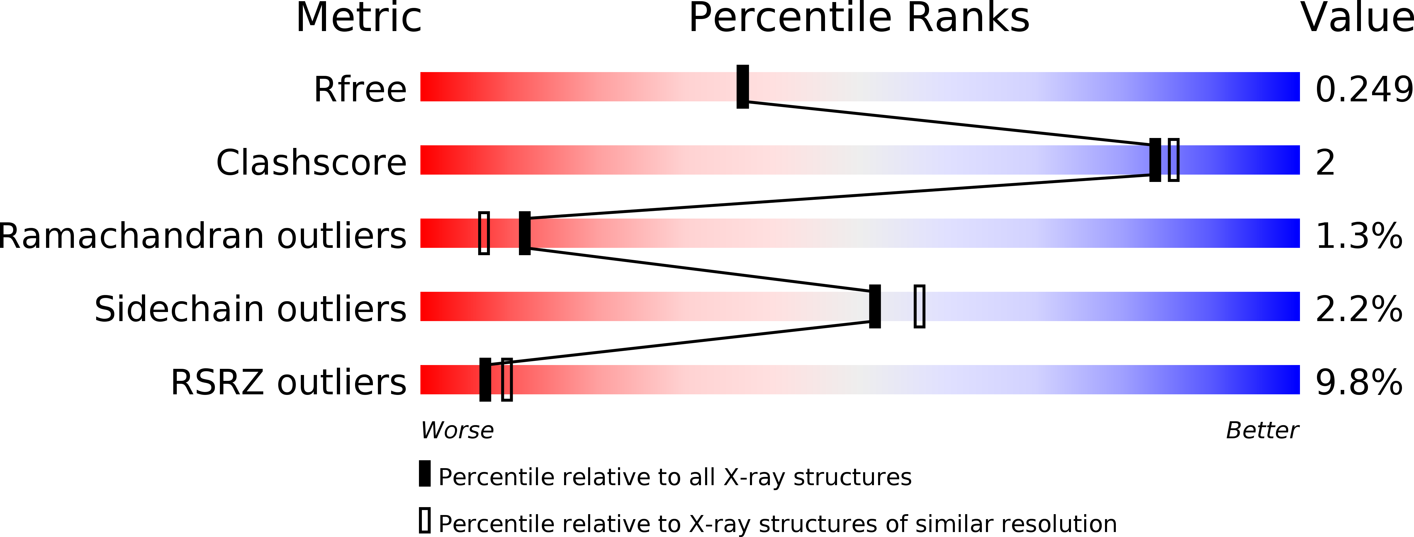
Resolution:
2.10 Å
R-Value Free:
0.23
R-Value Work:
0.17
R-Value Observed:
0.18
Space Group:
P 31 2 1
Macromolecular Entities
Polymer Type:polypeptide(L)
Molecule:Retinoic acid receptor RXR-alpha
Gene (Uniprot):RXRA
Chain IDs:A, B
Chain Length:87
Number of Molecules:2
Biological Source:Homo sapiens
Polymer Type:polydeoxyribonucleotide
Molecule:DNA (5'-D(*CP*TP*GP*GP*GP*TP*CP*AP*AP*AP*GP*TP*TP*CP*AP*TP*C)-3')
Chain IDs:C
Chain Length:17
Number of Molecules:1
Biological Source:Homo sapiens
Polymer Type:polydeoxyribonucleotide
Molecule:DNA (5'-D(*GP*AP*TP*GP*AP*AP*CP*TP*TP*TP*GP*AP*CP*CP*CP*AP*G)-3')
Chain IDs:D
Chain Length:17
Number of Molecules:1
Biological Source:Homo sapiens
Primary Citation
Modulation of RXR-DNA complex assembly by DNA context.
Mol. Cell. Endocrinol. 481 44 52 (2019)
PMID: 30476562 DOI: 10.1016/j.mce.2018.11.008

Abstact

Retinoid X Receptors (RXRs) act as dimer partners for several nuclear receptors including itself, binding to genomic DNA response elements and regulating gene transcription with cell and gene specificity. As homodimers, RXRs bind direct repeats of the half-site (A/G)G(G/T)TCA separated by 1 nucleotide (DR1) and little variability of this consensus site is observed for natural DR1s. However, these variations are responsible of the modulation of RXR receptors function through differential binding affinity and conformational changes. To further our understanding of the molecular mechanisms underlying RXR-DNA interactions, we examined how RXR DBDs bind to different DR1s using thermodynamics, X-ray crystallography and nuclear magnetic resonance (NMR) spectroscopy. We show that the half-site sequences modulate the binding cooperativity that results from the protein-protein contacts between the two DBDs. Chemical shifts perturbation NMR experiments revealed that sequence variations in half-sites induce changes that propagate from the protein-DNA interface to the dimerization interface throughout the DBD fold.

Legend

Protein

Chemical

Disease

Primary Citation of related structures