
Deposition Date
2017-12-15
Release Date
2018-11-28
Last Version Date
2024-05-08
Entry Detail
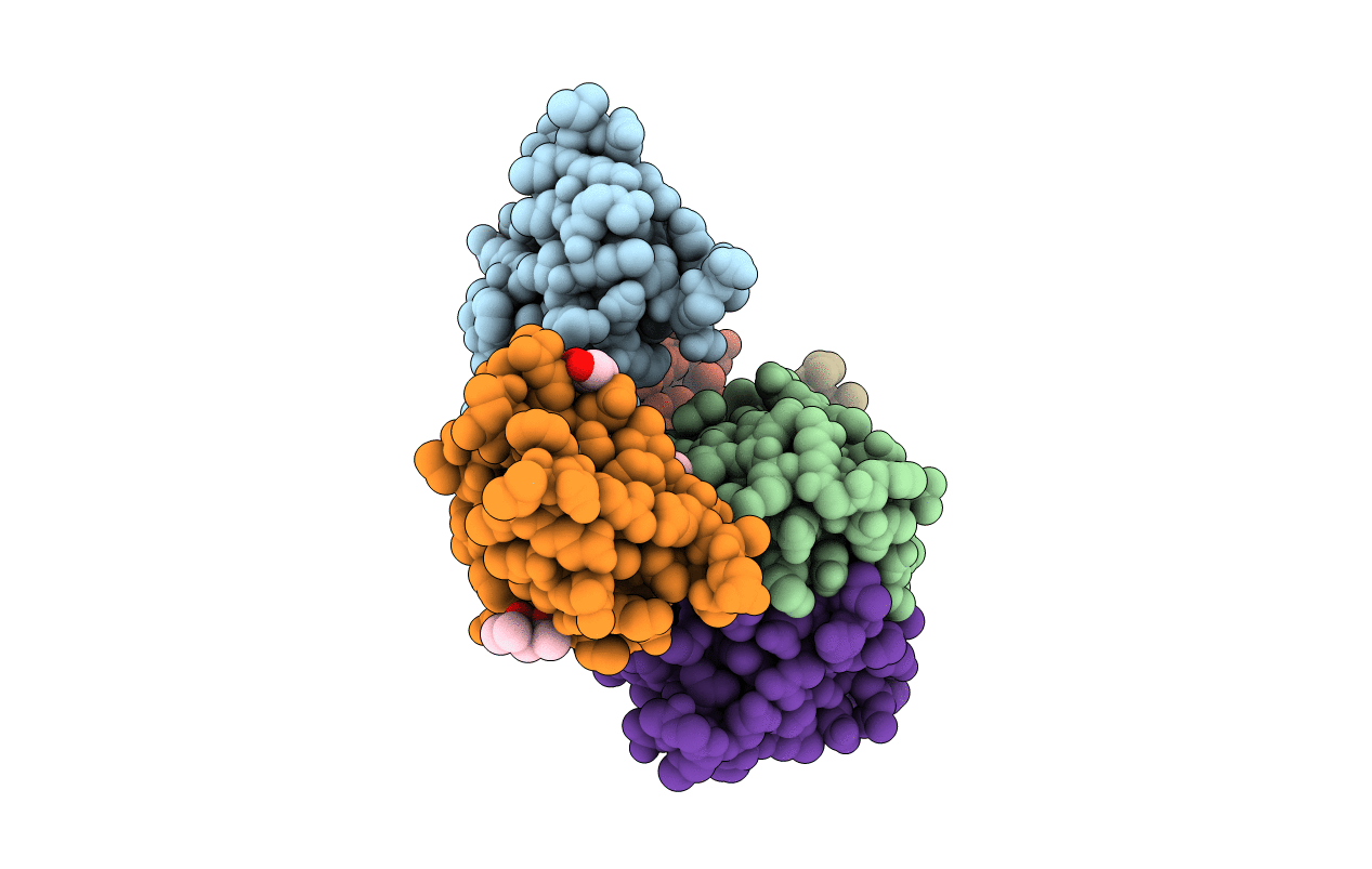
PDB ID:
6FAN
Keywords:
Title:
Crystal structure of putative CooT from Carboxydothermus hydrogenoformans
Biological Source:
Source Organism(s):
Expression System(s):
Method Details:
Experimental Method:
Resolution:
2.00 Å
R-Value Free:
0.27
R-Value Work:
0.23
R-Value Observed:
0.23
Space Group:
P 1 21 1
