Deposition Date
2017-12-14
Release Date
2018-01-31
Last Version Date
2024-05-15
Entry Detail
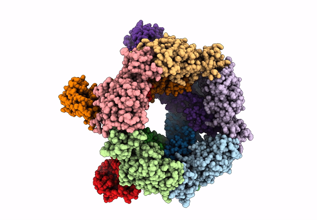
Biological Source:
Source Organism(s):
Rift valley fever virus (Taxon ID: 11588)
Expression System(s):
Method Details:
Experimental Method:
Resolution:
13.30 Å
Aggregation State:
PARTICLE
Reconstruction Method:
SINGLE PARTICLE