6F88 image
Entry Detail
PDB ID:
6F88
Keywords:
Title:
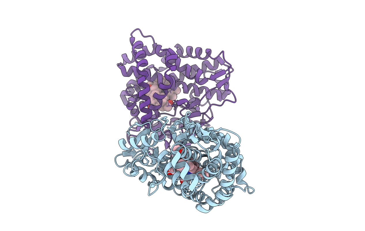
Crystal structure of cytochrome P450 CYP260A1 (S276N) bound with progesterone
Biological Source:
PDB Version:
Deposition Date:
2017-12-12
Release Date:
2018-03-14
Method Details:
Experimental Method:
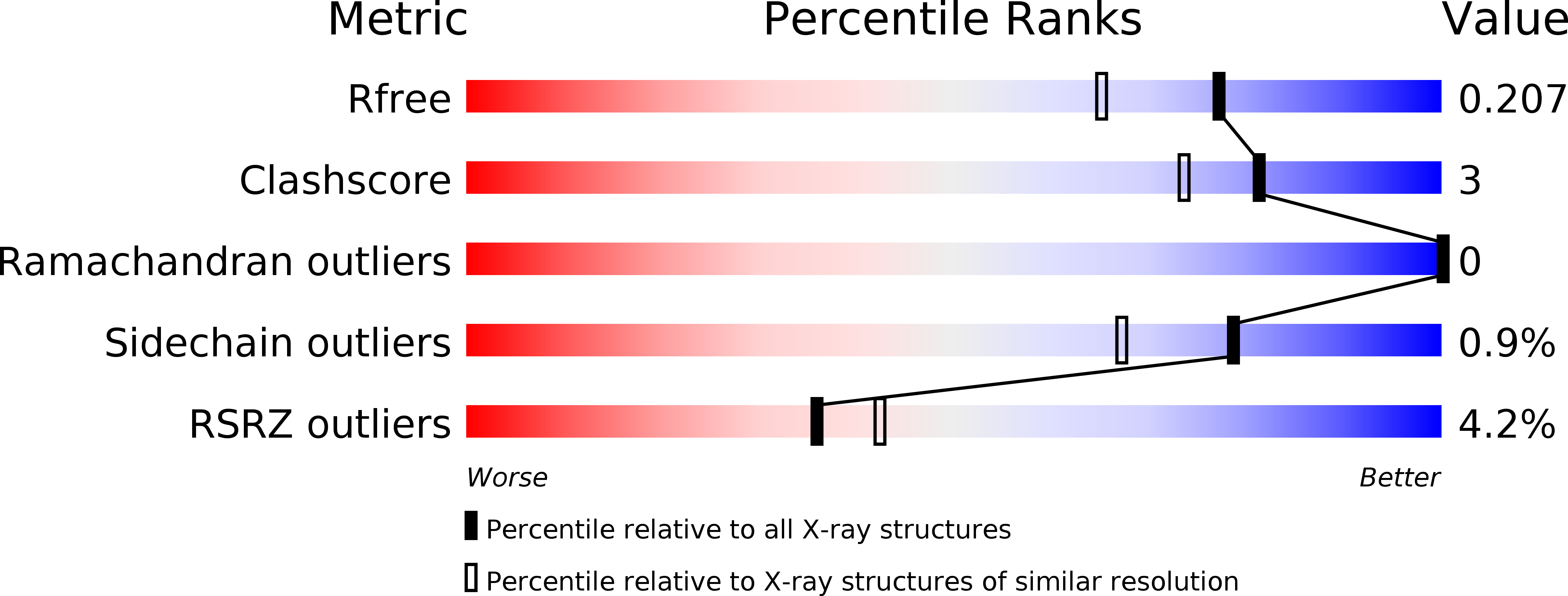
Resolution:
1.75 Å
R-Value Free:
0.20
R-Value Work:
0.16
R-Value Observed:
0.16
Space Group:
C 1 2 1
Macromolecular Entities
Polymer Type:polypeptide(L)
Description:Cytochrome P450 CYP260A1
Mutations:S276N
Chain IDs:A, B
Chain Length:400
Number of Molecules:2
Biological Source:Sorangium cellulosum (strain So ce56)
Primary Citation
Structure-Based Engineering of Steroidogenic CYP260A1 for Stereo- and Regioselective Hydroxylation of Progesterone.
ACS Chem. Biol. 13 1021 1028 (2018)
PMID: 29509407 DOI: 10.1021/acschembio.8b00026

Abstact

The production of regio- and stereoselectively hydroxylated steroids is of high pharmaceutical interest and can be achieved by cytochrome P450-based biocatalysts. CYP260A1 from Sorangium cellulosum strain So ce56 catalyzes hydroxylation of C19 or C21 steroids at the very unique 1α-position. However, the conversion of progesterone (PROG) by CYP260A1 is very unselective. In order to improve its selectivity we applied a semirational protein engineering approach, resulting in two different, highly regio- and stereoselective mutants by replacing a single serine residue (S276) of the substrate recognition site 5 with an asparagine or isoleucine. The S276N mutant converted PROG predominantly into 1α-hydroxy-PROG, while the S276I mutant led to 17α-hydroxy-PROG. We solved the high-resolution crystal structures of the PROG-bound S276N and S276I mutants, which revealed two different binding modes of PROG in the active site. The orientations were consistent with the exclusive 1α- (pro-1α binding mode) and 17α-hydroxylation (pro-17α-binding mode) of S276N and S276I, respectively. We observed that water-mediated hydrogen bonds contribute to the stabilization of the polar C3 and C17 substituents of PROG. Both binding modes of PROG may be stabilized in the wild-type enzyme. The change in regioselectivity is mainly driven by destabilizing the alternative binding mode due to steric hindrance and hydrogen bond disruption, caused by the mutations of Ser276. Thus, for the first time, the change in the selectivity of cytochrome P450-mediated steroid hydroxylation created by rational mutagenesis can be explained by the obtained 3D structures of the substrate-bound mutants, providing the basis for further experiments to engineer the biocatalyst toward novel steroid hydroxylation positions.

Legend

Protein

Chemical

Disease

Primary Citation of related structures