Deposition Date
2017-12-12
Release Date
2018-05-30
Last Version Date
2024-01-17
Entry Detail
PDB ID:
6F7X
Keywords:
Title:
Crystal structure of dimethylated RSL - cucurbit[7]uril complex, F432 form
Biological Source:
Source Organism(s):
Ralstonia solanacearum (Taxon ID: 305)
Expression System(s):
Method Details:
Experimental Method:
Resolution:
2.42 Å
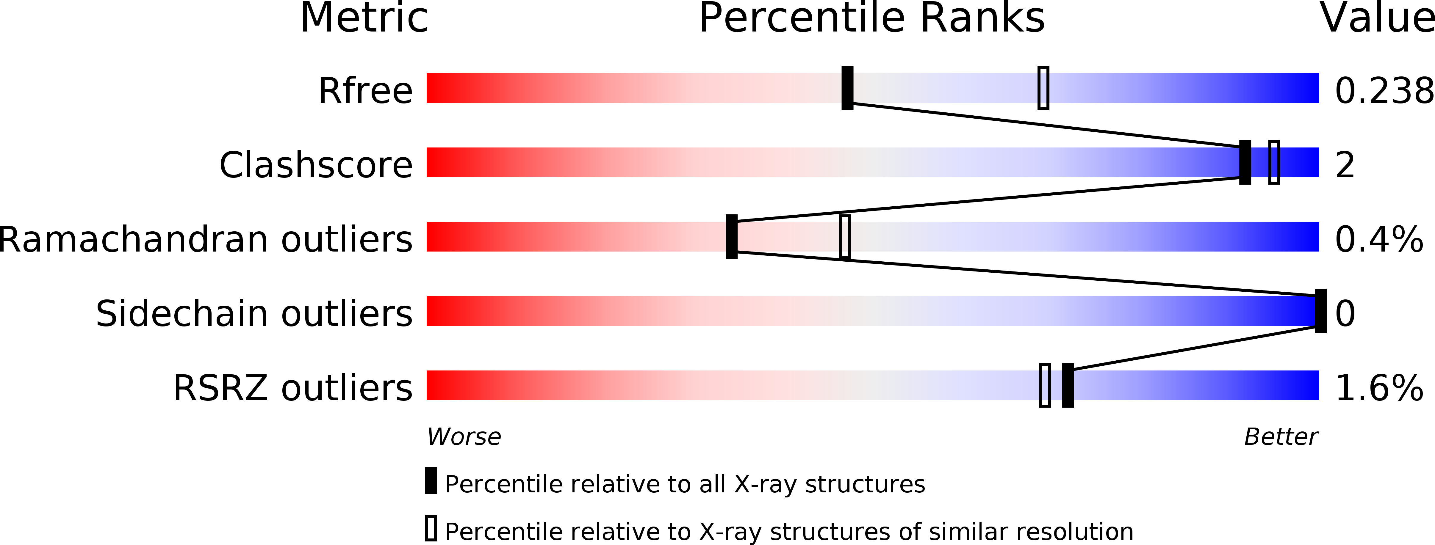
R-Value Free:
0.23
R-Value Work:
0.17
R-Value Observed:
0.17
Space Group:
F 4 3 2