Deposition Date
2017-12-11
Release Date
2018-10-31
Last Version Date
2024-11-20
Entry Detail
PDB ID:
6F7T
Keywords:
Title:
Crystal Structure of an Fab fragment in complex with a peptide from Bacillus subtilis RNase Y
Biological Source:
Source Organism(s):
Bacillus subtilis (Taxon ID: 1423)
Mus musculus (Taxon ID: 10090)
Mus musculus (Taxon ID: 10090)
Method Details:
Experimental Method:
Resolution:
2.60 Å
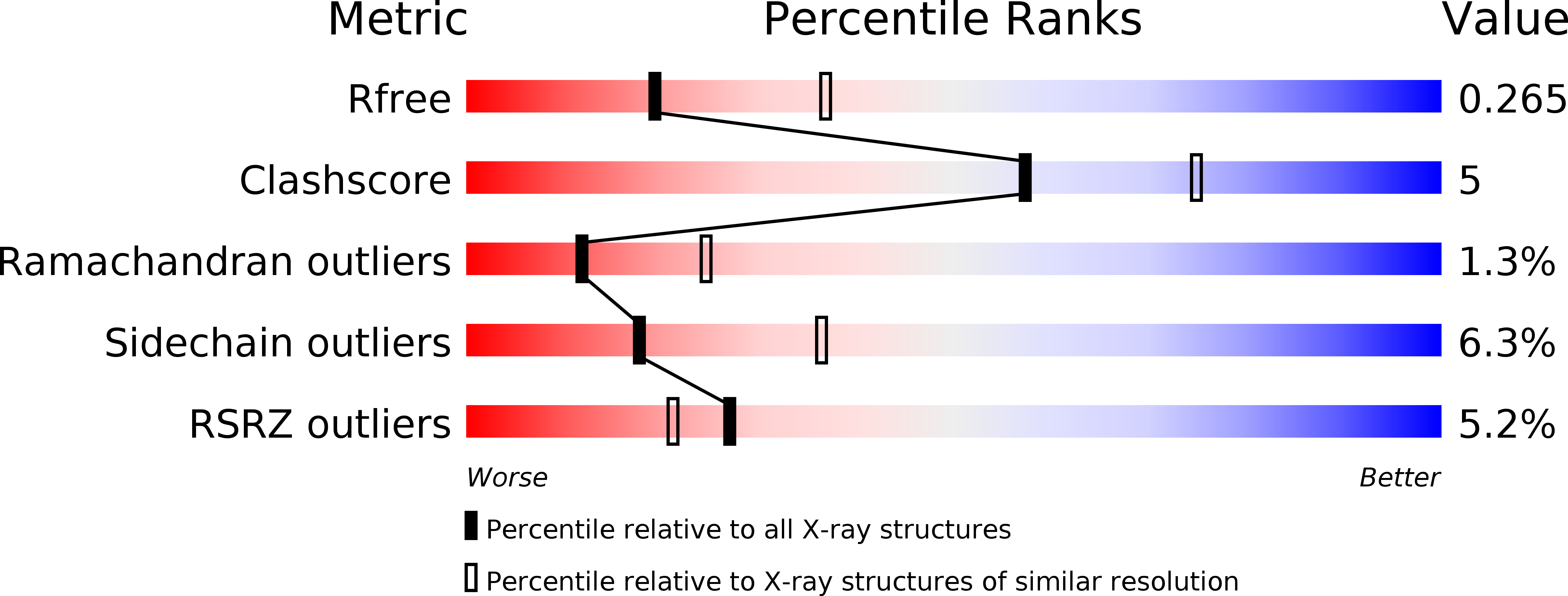
R-Value Free:
0.25
R-Value Work:
0.20
R-Value Observed:
0.20
Space Group:
P 21 21 2