
Deposition Date
2017-12-01
Release Date
2018-12-12
Last Version Date
2024-01-17
Entry Detail
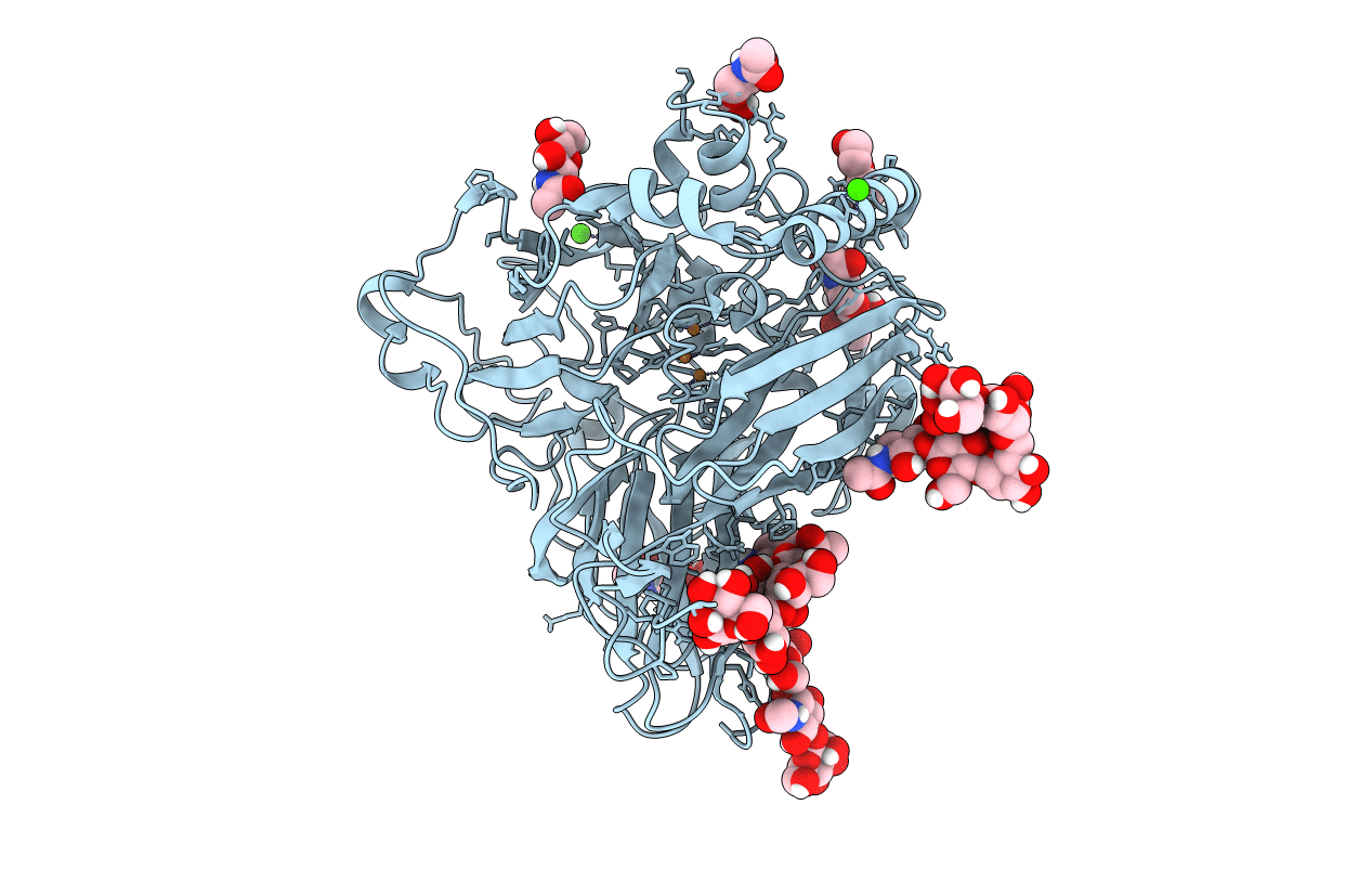
PDB ID:
6F5K
Keywords:
Title:
Crystal structure of laccase from Myceliophthora thermophila
Biological Source:
Source Organism(s):
Myceliophthora thermophila (Taxon ID: 78579)
Expression System(s):
Method Details:
Experimental Method:
Resolution:
1.62 Å
R-Value Free:
0.18
R-Value Work:
0.15
R-Value Observed:
0.15
Space Group:
C 2 2 21
