6F55 image
Deposition Date 2017-11-30
Release Date 2018-10-03
Last Version Date 2024-05-15
Entry Detail
PDB ID:
6F55
Title:
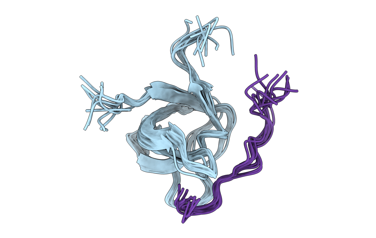
Complex structure of PACSIN SH3 domain and TRPV4 proline rich region
Biological Source:
Source Organism:
Gallus gallus (Taxon ID: 9031)
Homo sapiens (Taxon ID: 9606)
Host Organism:
Method Details:
Experimental Method:
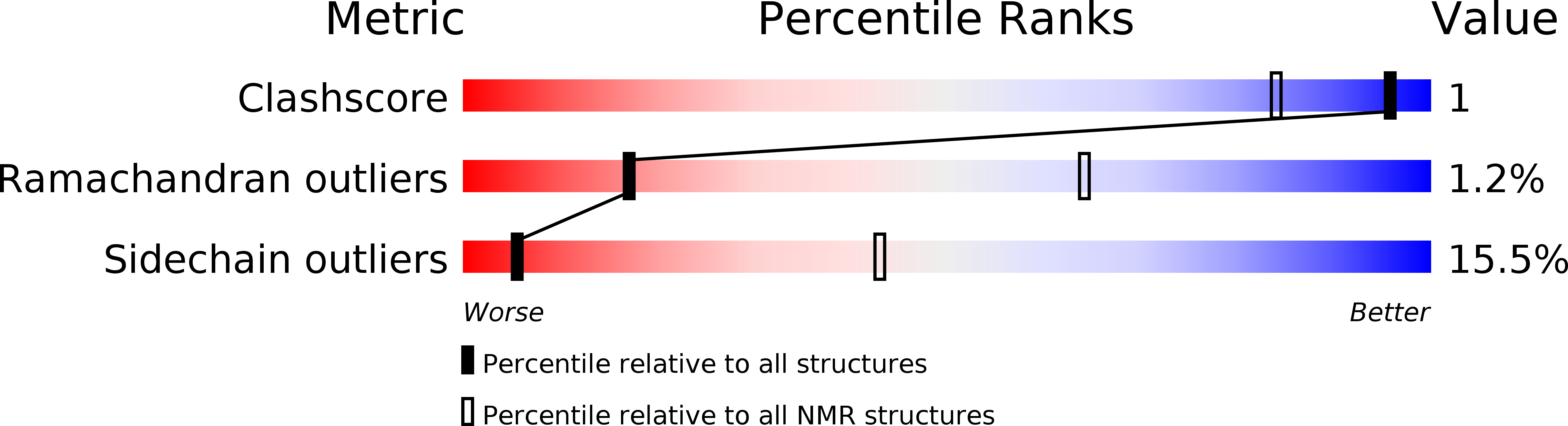
Conformers Calculated:
100
Conformers Submitted:
10
Selection Criteria:
structures with the least restraint violations
Macromolecular Entities
Structures with similar UniProt ID
Protein Blast
Polymer Type:polypeptide(L)
Molecule:PACSIN 3
Gene (Uniprot):PACSIN3
Chain IDs:A
Chain Length:67
Number of Molecules:1
Biological Source:Gallus gallus
Structures with similar UniProt ID
Protein Blast
Polymer Type:polypeptide(L)
Molecule:PRR
Chain IDs:B
Chain Length:15
Number of Molecules:1
Biological Source:Homo sapiens
Ligand Molecules
Primary Citation
Structural Basis of TRPV4 N Terminus Interaction with Syndapin/PACSIN1-3 and PIP2.
Structure 26 1583 1593.e5 (2018)
PMID: 30244966 DOI: 10.1016/j.str.2018.08.002

Abstact

Transient receptor potential (TRP) channels are polymodally regulated ion channels. TRPV4 (vanilloid 4) is sensitized by PIP2 and desensitized by Syndapin3/PACSIN3, which bind to the structurally uncharacterized TRPV4 N terminus. We determined the nuclear magnetic resonance structure of the Syndapin3/PACSIN3 SH3 domain in complex with the TRPV4 N-terminal proline-rich region (PRR), which binds as a class I polyproline II (PPII) helix. This PPII conformation is broken by a conserved proline in a cis conformation. Beyond the PPII, we find that the proximal TRPV4 N terminus is unstructured, a feature conserved across species thus explaining the difficulties in resolving it in previous structural studies. Syndapin/PACSIN SH3 domain binding leads to rigidification of both the PRR and the adjacent PIP2 binding site. We determined the affinities of the TRPV4 N terminus for PACSIN1, 2, and 3 SH3 domains and PIP2 and deduce a hierarchical interaction network where Syndapin/PACSIN binding influences the PIP2 binding site but not vice versa.

Legend

Protein

Chemical

Disease

Primary Citation of related structures